. 2010 Jan-Feb;5(1):1-8.
doi: 10.1002/cmmi.360.

Fluorescence lifetime imaging of activatable target specific molecular probes

Affiliations

Fluorescence lifetime imaging of activatable target specific molecular probes

Raphael Alford et al. Contrast Media Mol Imaging. 2010 Jan-Feb.

Abstract

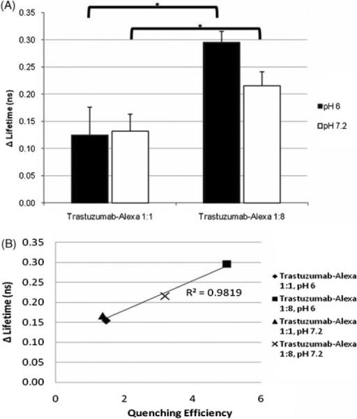
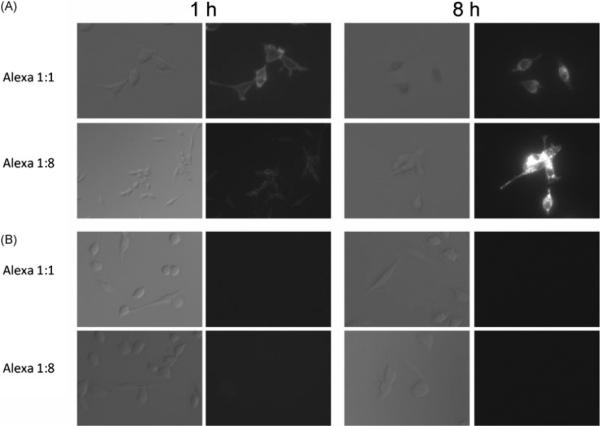
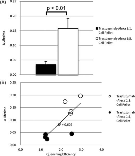
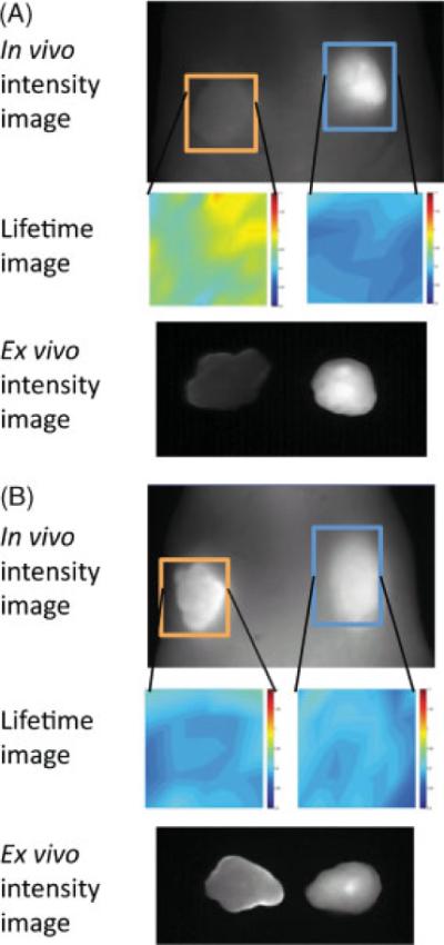
In vivo optical imaging using fluorescently labeled self-quenched monoclonal antibodies, activated through binding and internalization within target cells, results in excellent target-to-background ratios. We hypothesized that these molecular probes could be utilized to accurately report on cellular internalization with fluorescence lifetime imaging (FLI). Two imaging probes were synthesized, consisting of the antibody trastuzumab (targeting HER2/neu) conjugated to Alexa Fluor750 in ratios of either 1:8 or 1:1. Fluorescence intensity and lifetime of each conjugate were initially determined at endosomal pHs. Since the 1:8 conjugate is self-quenched, the fluorescence lifetime of each probe was also determined after exposure to the known dequencher SDS. In vitro imaging experiments were performed using 3T3/HER2(+) and BALB/3T3 (HER2(-)) cell lines. Changes in fluorescence lifetime correlated with temperature- and time-dependent cellular internalization. In vivo imaging studies in mice with dual flank tumors [3T3/HER2(+) and BALB/3T3 (HER2(-))] detected a minimal difference in FLI. In conclusion, fluorescence lifetime imaging monitors the internalization of target-specific activatable antibody-fluorophore conjugates in vitro. Challenges remain in adapting this methodology to in vivo imaging.


Figures

Figure 1
Figure 1
(A) Trastuzumab–Alexa 1:8 demonstrates significant (p < 0.01) dequenching of fluorescence intensity with the addition of SDS, whereas trastuzumab–Alexa 1:1 demonstrates a significant change (p < 0.05) only at the lower pH. (B) Trastuzumab–Alexa 1:8 demonstrates a significantly greater quenching efficiency (quenching efficiency = dequenched conjugate fluorescence intensity/quenched conjugate fluorescence intensity) than trastuzumab–Alexa 1:1 conjugate; n = 4.
Figure 2
Figure 2
(A) Trastuzumab–Alexa 1:8 demonstrates a larger increase in lifetime with dequenching. (B) A correlation is noted between larger quenching efficiencies and larger lifetime increases; n = 3.
Figure 3
Figure 3
Serial DIC and fluorescence microscopy studies. (A) 3T3/HER2+ cells were incubated at 10 μg/ml of either traztuzumab–Alexa 1:1, or traztuzumab–Alexa 1:8 for 1 or 8 h. The fluorescence signal is distributed uniformly on cell surface after 1 h, and can be seen within the cell after 8 h. Self-quenched conjugated demonstrates a higher fluorescence signal following internalization. (B) Conjugates were incubated in BALB/3T3 (HER-2) cells. No fluorophores appeared to be bound or internalized by the cells. Magnification: 400×. DIC exposure time: 500 μs. Fluorescence exposure time: 100 ms.
Figure 4
Figure 4
(A) Demonstrates the significantly greater increase (p < 0.01) in lifetime secondary to cellular internalization by HER2+ cells; n=4. (B). A positive correlation between quenching efficiencies and lifetime is again observed.
Figure 5
Figure 5
Black and white images are in vivo spectral fluorescence images showing dorsum of tumor-bearing mice and ex vivo resected tumors, unmixed to select for Alexa Fluor 750. The orange box represents the HER-2+ region of interest (ROI), and the blue box represents the HER-2- ROI. Lifetime images corresponding to each ROI are directly beneath the ROI. Lifetimes are displayed on a pseudocolor scale, ranging from 0.8 to 1.1 ns. (A) The one mouse with increase in lifetime in the HER-2+ tumor as opposed to the HER-2; (B) no difference between the HER-2+ and HER-2- tumors, which was the more consistent finding.

Similar articles

Cited by

References

    1. Massoud TF, Gambhir SS. Molecular imaging in living subjects: seeing fundamental biological processes in a new light. Genes Dev. 2003;17:545–580. - PubMed
    1. Ntziachristos V. Fluorescence molecular imaging. Annu Rev Biomed Eng. 2006;8:1–33. - PubMed
    1. Weissleder R. Molecular imaging in cancer. Science. 2006;312:1168–1171. - PubMed
    1. Ogawa M, Kosaka N, Longmire MR, Urano Y, Choyke PL, Kobayashi H. Fluorophore-quencher based activatable targeted optical probes for detecting in vivo cancer metastases. Mol Pharm. 2009;6:386–395. - PMC - PubMed
    1. Ogawa M, Regino CA, Choyke PL, Kobayashi H. In vivo target-specific activatable near-infrared optical labeling of humanized monoclonal antibodies. Mol Cancer Ther. 2009;8:232–239. - PMC - PubMed

Publication types