Review
. 2010 Jan 28;16(4):411-7.
doi: 10.3748/wjg.v16.i4.411.

Endoscope-guided pneumatic dilation for treatment of esophageal achalasia

Review

Endoscope-guided pneumatic dilation for treatment of esophageal achalasia

Seng-Kee Chuah et al. World J Gastroenterol. .

Abstract

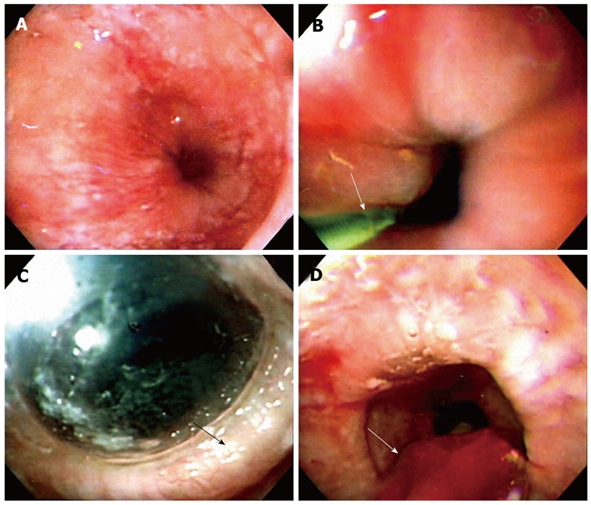
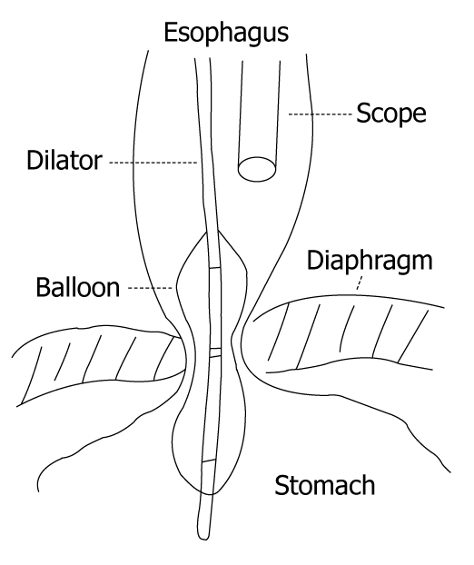
Pneumatic dilation (PD) is considered to be the first line nonsurgical therapy for achalasia. The principle of the procedure is to weaken the lower esophageal sphincter by tearing its muscle fibers by generating radial force. The endoscope-guided procedure is done without fluoroscopic control. Clinicians usually use a low-compliance balloon such as Rigiflex dilator to perform endoscope-guided PD for the treatment of esophageal achalasia. It has the advantage of determining mucosal injury during the dilation process, so that a repeat endoscopy is not needed to assess the mucosal tearing. Previous studies have shown that endoscope-guided PD is an efficient and safe nonsurgical therapy with results that compare well with other treatment modalities. Although the results may be promising, long-term follow-up is required in the near future.


Figures

Figure 1
Figure 1
Technique of endoscope-guided pneumatic dilation (PD). A: A dilated low esophageal lumen with tight gastroesophageal junction under endoscope-guided PD; B: The endoscope is inserted down to the duodenum. A guide-wire (arrow) is placed into the duodenum under endoscopic guidance and the endoscope is removed. A Regiflex balloon dilator, which is marked with a thick colored marker at the mid-section of the balloon, is passed over the guide wire to the stomach; C: The endoscope is reinserted to serve as a guide to control the position of the balloon in the esophagus. The balloon is withdrawn to the esophagus, until the mark reaches the gastroesophageal junction. The balloon is then inflated up to 12 psi and maintained for 60 s, until an ischemic ring (arrow) at the LES is seen by the endoscope through the transparent balloon; D: The balloon is flattened completely and removed together with the endoscope (arrow).
Figure 2
Figure 2
Almost all endoscope-guided PD are performed by using a low compliance Rigiflex balloon dilator by positioning the endoscope above the balloon without fluoroscopy. The advantages are that the procedure can be performed without any extra equipment in a time and cost-effective manner.

References

    1. Chuah SK, Kuo CM, Wu KL, Changchien CS, Hu TH, Wang CC, Chiu YC, Chou YP, Hsu PI, Chiu KW, et al. Pseudoachalasia in a patient after truncal vagotomy surgery successfully treated by subsequent pneumatic dilations. World J Gastroenterol. 2006;12:5087–5090. - PMC - PubMed
    1. Gockel I, Eckardt VF, Schmitt T, Junginger T. Pseudoachalasia: a case series and analysis of the literature. Scand J Gastroenterol. 2005;40:378–385. - PubMed
    1. Mohamed A. Education and Imaging. Gastrointestinal: pseudoachalasia caused by a lower esophageal stromal tumor. J Gastroenterol Hepatol. 2009;24:1152. - PubMed
    1. Saino G, Bona D, Nencioni M, Rubino B, Bonavina L. Laparoscopic diagnosis of pleural mesothelioma presenting with pseudoachalasia. World J Gastroenterol. 2009;15:3569–3572. - PMC - PubMed
    1. Moorman AJ, Oelschlager BK, Rulyak SJ. Pseudoachalasia caused by retroperitoneal B-cell lymphoma. Clin Gastroenterol Hepatol. 2008;6:A32. - PubMed

Substances