Percutaneous catheter drainage in combination with choledochoscope-guided debridement in treatment of peripancreatic infection
- PMID: 20101781
- PMCID: PMC2811808
- DOI: 10.3748/wjg.v16.i4.513
Percutaneous catheter drainage in combination with choledochoscope-guided debridement in treatment of peripancreatic infection
Abstract
Aim: To introduce and evaluate the new method used in treatment of pancreatic and peripancreatic infections secondary to severe acute pancreatitis (SAP).
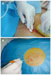
Methods: A total of 42 SAP patients initially underwent ultrasound-guided percutaneous puncture and catheterization. An 8-Fr drainage catheter was used to drain the infected peripancreatic necrotic foci for 3-5 d. The sinus tract of the drainage catheter was expanded gradually with a skin expander, and the 8-Fr drainage catheter was replaced with a 22-Fr drainage tube after 7-10 d. Choledochoscope-guided debridement was performed repeatedly until the infected peripancreatic tissue was effectively removed through the drainage sinus tract.
Results: Among the 42 patients, the infected peripancreatic tissue or abscess was completely removed from 38 patients and elective cyst-jejunum anastomosis was performed in 4 patients due to formation of pancreatic pseudocysts. No death and complication occurred during the procedure.
Conclusion: Percutaneous catheter drainage in combination with choledochoscope-guided debridement is a simple, safe and reliable treatment procedure for peripancreatic infections secondary to SAP.
Figures


Comment in
-
Comments on the article about the treatment of peripancreatic infection.World J Gastroenterol. 2010 May 14;16(18):2321-2. doi: 10.3748/wjg.v16.i18.2321. World J Gastroenterol. 2010. PMID: 20458775 Free PMC article.
References
-
- Beger HG, Rau B, Mayer J, Pralle U. Natural course of acute pancreatitis. World J Surg. 1997;21:130–135. - PubMed
-
- Werner J, Hartwig W, Hackert T, Büchler MW. Surgery in the treatment of acute pancreatitis--open pancreatic necrosectomy. Scand J Surg. 2005;94:130–134. - PubMed
-
- Banks PA, Freeman ML. Practice guidelines in acute pancreatitis. Am J Gastroenterol. 2006;101:2379–2400. - PubMed
-
- Balthazar EJ. Complications of acute pancreatitis: clinical and CT evaluation. Radiol Clin North Am. 2002;40:1211–1227. - PubMed
-
- Bouvet M, Moussa AR. Pancreatic abscess. In: Cameron JL, editor. Current surgical therapy. 8th ed. Philadelphia: Elsevier Mosby; 2004. pp. 476–480.
Publication types
MeSH terms
LinkOut - more resources
Full Text Sources
Miscellaneous

