

A perivascular epithelioid cell tumor of the stomach: an unsuspected diagnosis
- PMID: 20101783
- PMCID: PMC2811810
- DOI: 10.3748/wjg.v16.i4.522
A perivascular epithelioid cell tumor of the stomach: an unsuspected diagnosis
Abstract
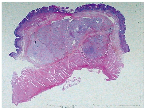
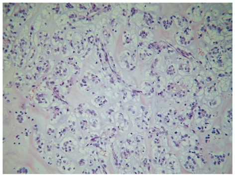
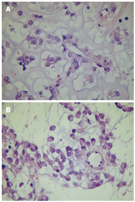
Perivascular epithelioid cell tumor (PEComa) is a rare mesenchymal neoplasia and currently well recognized as a distinct entity with characteristic morphological, immunohistochemical and molecular findings. We report a case of PEComa arising in the antrum of a 71-year-old female with melena. The tumor, located predominantly in the submucosa as a well delimited nodule, measured 3.0 cm in diameter and was completely resected, with no evidence of the disease elsewhere. Histologically, it was composed predominantly of eosinophilic epithelioid cells arranged in small nests commonly related to variably sized vessels, with abundant extracellular material, moderate nuclear variation and discrete mitotic activity. No necrosis, angiolymphatic invasion or perineural infiltration was seen. Tumor cells were uniformly positive for vimentin, smooth muscle actin, desmin and melan A. Although unusual, PEComa should be considered in the differential diagnosis of gastric neoplasia with characteristic epithelioid and oncocytic features and prominent vasculature.
Figures
References
-
- Folpe AL. Neoplasms with perivascular epithelioid cell differentiation (PEComas) In: Fletcher CDM, Unni KK, Mertens F, editors. World Health Organization Classification of Tumors. Pathology and Genetics of Tumors of Soft Tissue and Bone. Lyon: IARC Press; 2002. pp. 221–222.
-
- Hornick JL, Fletcher CD. PEComa: what do we know so far? Histopathology. 2006;48:75–82. - PubMed
-
- Agaimy A, Wünsch PH. Perivascular epithelioid cell sarcoma (malignant PEComa) of the ileum. Pathol Res Pract. 2006;202:37–41. - PubMed
-
- Baek JH, Chung MG, Jung DH, Oh JH. Perivascular epithelioid cell tumor (PEComa) in the transverse colon of an adolescent: a case report. Tumori. 2007;93:106–108. - PubMed
Publication types
MeSH terms
LinkOut - more resources
Full Text Sources
Medical
