

Two-step synthesis of galactosylated human serum albumin as a targeted optical imaging agent for peritoneal carcinomatosis
- PMID: 20102220
- PMCID: PMC3230036
- DOI: 10.1021/jm901228u
Two-step synthesis of galactosylated human serum albumin as a targeted optical imaging agent for peritoneal carcinomatosis
Abstract
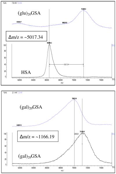
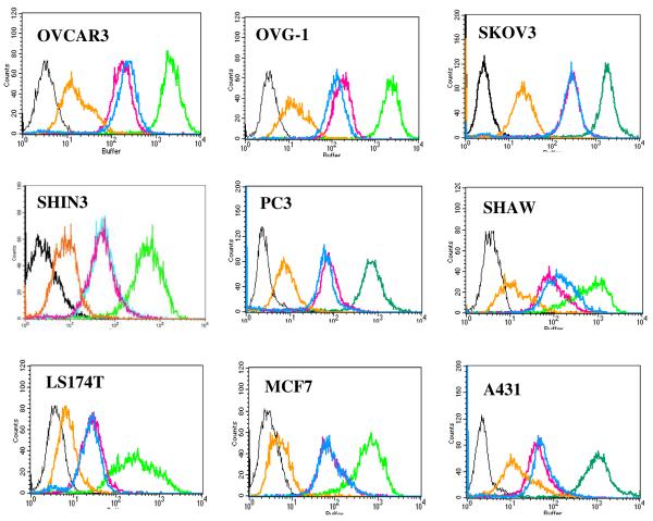
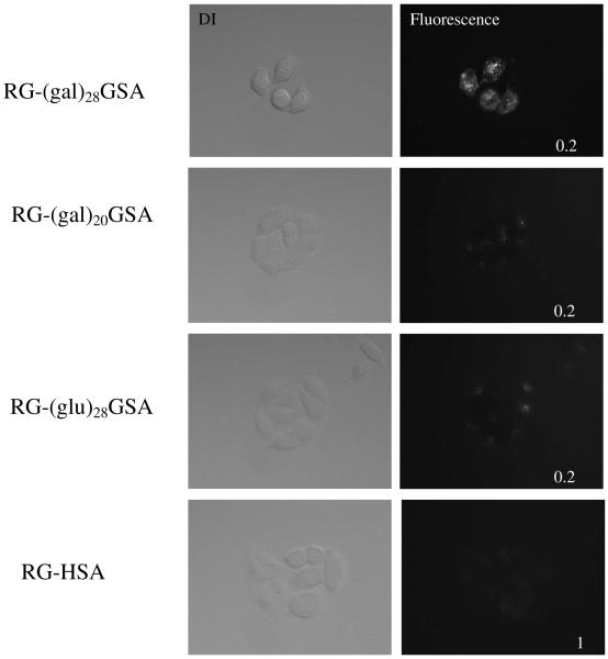
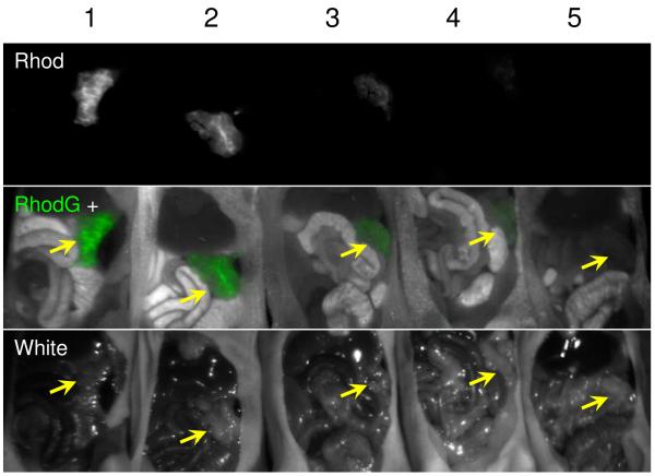
An optical probe, RG-(gal)(28)GSA, was synthesized to improve the detection of peritoneal implants by targeting the beta-d-galactose receptors highly expressed on the cell surface of a wide variety of cancers arising from the ovary, pancreas, colon, and stomach. Evaluation of RG-(gal)(28)GSA, RG-(gal)(20)GSA, glucose-analogue RG-(glu)(28)GSA, and control RG-HSA demonstrates specificity for the galactose, binding to several human adenocarcinoma cell lines, and cellular internalization. Studies using peritoneally disseminated SHIN3 xenografts in mice also confirmed a preference for galactose with the ability to detect submillimeter size lesions. Preliminary toxicity study for RG-(gal)(28)GSA using Balb/c mice reveal no toxic effects up to 100x of the standard imaging dose of 1 mg/kg administered either intraperitoneally or intravenously. These data indicate that RG-(gal)(28)GSA can selectively target a variety of human adenocarcinomas, can improve intraoperative or endoscopic tumor detection and resection, and may have little or no toxic in vivo effects; hence, it may be clinically translatable.
Figures
Similar articles
-
Galactosyl human serum albumin-NMP1 conjugate: a near infrared (NIR)-activatable fluorescence imaging agent to detect peritoneal ovarian cancer metastases.Bioconjug Chem. 2012 Aug 15;23(8):1671-9. doi: 10.1021/bc3002419. Epub 2012 Jul 30. Bioconjug Chem. 2012. PMID: 22799539 Free PMC article.
-
A self-quenched galactosamine-serum albumin-rhodamineX conjugate: a "smart" fluorescent molecular imaging probe synthesized with clinically applicable material for detecting peritoneal ovarian cancer metastases.Clin Cancer Res. 2007 Nov 1;13(21):6335-43. doi: 10.1158/1078-0432.CCR-07-1004. Clin Cancer Res. 2007. PMID: 17975145
-
D-galactose receptor-targeted in vivo spectral fluorescence imaging of peritoneal metastasis using galactosamin-conjugated serum albumin-rhodamine green.J Biomed Opt. 2007 Sep-Oct;12(5):051501. doi: 10.1117/1.2779351. J Biomed Opt. 2007. PMID: 17994865
-
Near-infrared photoimmunotherapy with galactosyl serum albumin in a model of diffuse peritoneal disseminated ovarian cancer.Oncotarget. 2016 Nov 29;7(48):79408-79416. doi: 10.18632/oncotarget.12710. Oncotarget. 2016. PMID: 27765903 Free PMC article.
-
Avidin conjugated to tetramethyl-6-carboxyrhodamine-QSY®7.2009 Feb 20 [updated 2009 Apr 6]. In: Molecular Imaging and Contrast Agent Database (MICAD) [Internet]. Bethesda (MD): National Center for Biotechnology Information (US); 2004–2013. 2009 Feb 20 [updated 2009 Apr 6]. In: Molecular Imaging and Contrast Agent Database (MICAD) [Internet]. Bethesda (MD): National Center for Biotechnology Information (US); 2004–2013. PMID: 20641900 Free Books & Documents. Review.
Cited by
-
A portable fluorescence camera for testing surgical specimens in the operating room: description and early evaluation.Mol Imaging Biol. 2011 Oct;13(5):862-7. doi: 10.1007/s11307-010-0438-2. Mol Imaging Biol. 2011. PMID: 20960235 Free PMC article.
-
In vivo kinetics and biodistribution analysis of neoglycoproteins: effects of chemically introduced glycans on proteins.Glycoconj J. 2014 May;31(4):273-9. doi: 10.1007/s10719-014-9520-3. Epub 2014 Apr 5. Glycoconj J. 2014. PMID: 24705934 Review.
-
Recent Advancements and Strategies for Overcoming the Blood-Brain Barrier Using Albumin-Based Drug Delivery Systems to Treat Brain Cancer, with a Focus on Glioblastoma.Polymers (Basel). 2023 Oct 2;15(19):3969. doi: 10.3390/polym15193969. Polymers (Basel). 2023. PMID: 37836018 Free PMC article. Review.
-
Galactosyl human serum albumin-NMP1 conjugate: a near infrared (NIR)-activatable fluorescence imaging agent to detect peritoneal ovarian cancer metastases.Bioconjug Chem. 2012 Aug 15;23(8):1671-9. doi: 10.1021/bc3002419. Epub 2012 Jul 30. Bioconjug Chem. 2012. PMID: 22799539 Free PMC article.
-
Analysis of carbohydrates and glycoconjugates by matrix-assisted laser desorption/ionization mass spectrometry: an update for 2009-2010.Mass Spectrom Rev. 2015 May-Jun;34(3):268-422. doi: 10.1002/mas.21411. Epub 2014 May 26. Mass Spectrom Rev. 2015. PMID: 24863367 Free PMC article. Review.
References
-
- Heron MP, Hoyert DL, Xu J, Scott C, Tejada-Vera B. Deaths: Preliminary data for 2006. National Vital Statistics Reports. 2008;56:1–52. - PubMed
-
- Jemal A, Siegel R, Ward E, Hao Y, Xu J, Murray T, Thun MJ. Cancer statistics, 2008. CA Cancer J Clin. 2008;58:71–96. - PubMed
-
- Hoffman RM. The multiple uses of fluorescent proteins to visualize cancer in vivo. Nat Rev Cancer. 2005;5:796–806. - PubMed
-
- Hoffman RM, Yang M. Subcellular imaging in the live mouse. Nat Protocols. 2006;1:775–782. - PubMed
-
- Hoffman RM, Yang M. Color-coded fluorescence imaging of tumor-host interactions. Nat Protocols. 2006;1:9–928. - PubMed
Publication types
MeSH terms
Substances
Grants and funding
LinkOut - more resources
Full Text Sources
Medical
