

Peptide-based probes for targeted molecular imaging
- PMID: 20102226
- PMCID: PMC2829605
- DOI: 10.1021/bi901135x
Peptide-based probes for targeted molecular imaging
Abstract
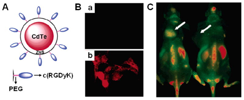
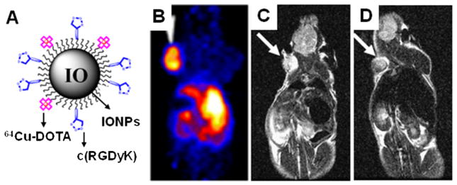
Targeted molecular imaging techniques have become indispensable tools in modern diagnostics because they provide accurate and specific diagnosis of disease information. Conventional nonspecific contrast agents suffer from low targeting efficiency; thus, the use of molecularly targeted imaging probes is needed depending on different imaging modalities. Although recent technologies have yielded various strategies for designing smart probes, utilization of peptide-based probes has been most successful. Phage display technology and combinatorial peptide chemistry have profoundly impacted the pool of available targeting peptides for the efficient and specific delivery of imaging labels. To date, selected peptides that target a variety of disease-related receptors and biomarkers are in place. These targeting peptides can be coupled with the appropriate imaging moieties or nanoplatforms on demand with the help of sophisticated bioconjugation or radiolabeling techniques. This review article examines the current trends in peptide-based imaging probes developed for in vivo applications. We discuss the advantage of and challenges in developing peptide-based probes and summarize current systems with respect to their unique design strategies and applications.
Figures
Similar articles
-
Target-specific delivery of peptide-based probes for PET imaging.Adv Drug Deliv Rev. 2010 Aug 30;62(11):1005-22. doi: 10.1016/j.addr.2010.09.004. Epub 2010 Sep 17. Adv Drug Deliv Rev. 2010. PMID: 20851156 Review.
-
Tumor-targeting peptides from combinatorial libraries.Adv Drug Deliv Rev. 2017 Feb;110-111:13-37. doi: 10.1016/j.addr.2016.05.009. Epub 2016 May 19. Adv Drug Deliv Rev. 2017. PMID: 27210583 Free PMC article. Review.
-
From combinatorial chemistry to cancer-targeting peptides.Mol Pharm. 2007 Sep-Oct;4(5):631-51. doi: 10.1021/mp700073y. Epub 2007 Sep 20. Mol Pharm. 2007. PMID: 17880166 Review.
-
Peptides for in vivo target-specific cancer imaging.Mini Rev Med Chem. 2010 Jan;10(1):87-97. doi: 10.2174/138955710791112596. Mini Rev Med Chem. 2010. PMID: 20380643 Review.
-
Peptide-receptor ligands and multivalent approach.Anticancer Agents Med Chem. 2012 Jun;12(5):416-27. doi: 10.2174/187152012800617849. Anticancer Agents Med Chem. 2012. PMID: 22292761 Review.
Cited by
-
Preliminary study of injury from heating systemically delivered, nontargeted dextran-superparamagnetic iron oxide nanoparticles in mice.Nanomedicine (Lond). 2012 Nov;7(11):1697-711. doi: 10.2217/nnm.12.65. Epub 2012 Jul 26. Nanomedicine (Lond). 2012. PMID: 22830502 Free PMC article.
-
Fluorogen activating protein-affibody probes: modular, no-wash measurement of epidermal growth factor receptors.Bioconjug Chem. 2015 Jan 21;26(1):137-44. doi: 10.1021/bc500525b. Epub 2014 Dec 19. Bioconjug Chem. 2015. PMID: 25490520 Free PMC article.
-
A TMVP1-modified near-infrared nanoprobe: molecular imaging for tumor metastasis in sentinel lymph node and targeted enhanced photothermal therapy.J Nanobiotechnology. 2023 Apr 17;21(1):130. doi: 10.1186/s12951-023-01883-6. J Nanobiotechnology. 2023. PMID: 37069646 Free PMC article.
-
Shining a Light on Venom-Peptide Receptors: Venom Peptides as Targeted Agents for In Vivo Molecular Imaging.Toxins (Basel). 2024 Jul 4;16(7):307. doi: 10.3390/toxins16070307. Toxins (Basel). 2024. PMID: 39057947 Free PMC article. Review.
-
Comparison of prostate-specific membrane antigen ligands in clinical translation research for diagnosis of prostate cancer.Cancer Rep (Hoboken). 2019 Aug;2(4):e1169. doi: 10.1002/cnr2.1169. Epub 2019 Apr 2. Cancer Rep (Hoboken). 2019. PMID: 32721116 Free PMC article. Review.
References
-
- Hoffman JM, Gambhir SS. Molecular imaging: the vision and opportunity for radiology in the future. Radiology. 2007;244:39–47. - PubMed
-
- Cai W, Chen X. Nanoplatforms for targeted molecular imaging in living subjects. Small. 2007;3:1840–1854. - PubMed
-
- Wu AM, Olafsen T. Antibodies for molecular imaging of cancer. Cancer J. 2008;14:191–197. - PubMed
-
- Aloj L, Morelli G. Design, synthesis and preclinical evaluation of radiolabeled peptides for diagnosis and therapy. Curr Pharm Des. 2004;10:3009–3031. - PubMed
-
- Reubi JC, Maecke HR. Peptide-based probes for cancer imaging. J Nucl Med. 2008;49:1735–1738. - PubMed
Publication types
MeSH terms
Substances
Grants and funding
LinkOut - more resources
Full Text Sources
Other Literature Sources
