. 2010 Mar 2;49(8):1568-76.
doi: 10.1021/bi9021878.

Binding of Munc18-1 to synaptobrevin and to the SNARE four-helix bundle

Affiliations

Binding of Munc18-1 to synaptobrevin and to the SNARE four-helix bundle

Yi Xu et al. Biochemistry. .

Abstract

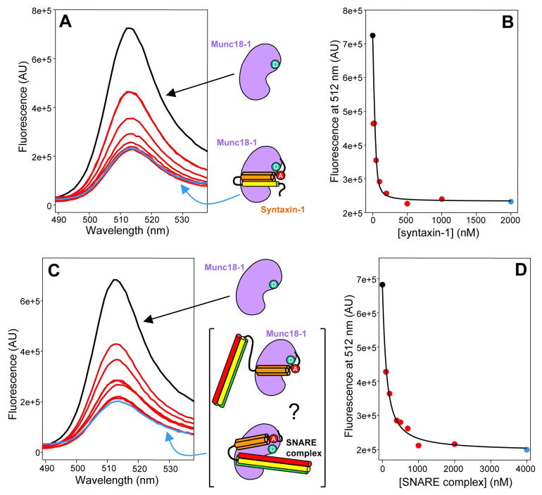
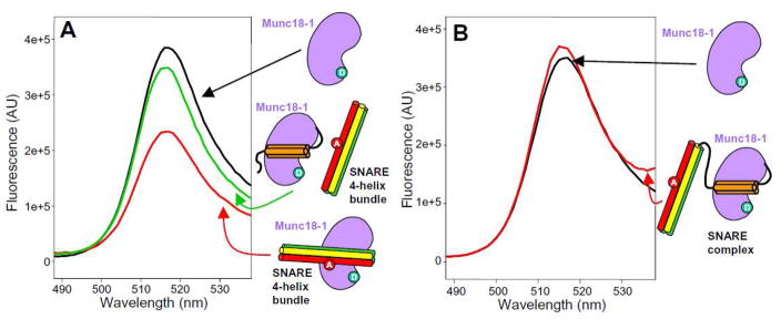
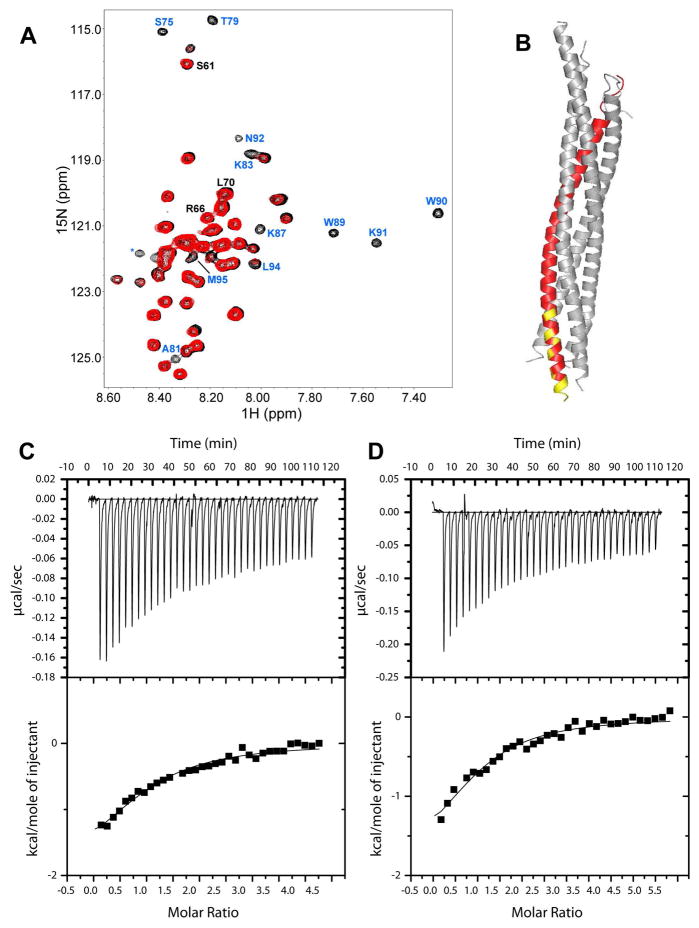
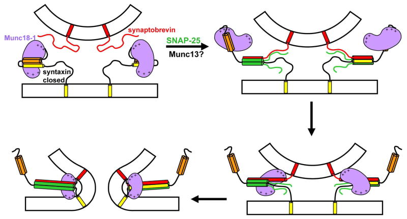
Sec1/Munc18 (SM) proteins and soluble N-ethylmaleimide sensitive factor attachment protein receptors (SNAREs) form part of the core intracellular membrane fusion machinery, but it is unclear how they cooperate in membrane fusion. The synaptic vesicle SNARE synaptobrevin and the plasma membrane SNAREs syntaxin-1 and SNAP-25 assemble into a tight SNARE complex that includes a four-helix bundle formed by their SNARE motifs and is key for fusion. The neuronal SM protein Munc18-1 binds to syntaxin-1 and to the SNARE complex through interactions with the syntaxin-1 N-terminal region that are critical for neurotransmitter release. It has been proposed that Munc18-1 also binds to synaptobrevin and to the SNARE four-helix bundle and that such interactions might be crucial for membrane fusion, but definitive, direct evidence of these interactions has not been described. Using diverse biophysical approaches, we now demonstrate that Munc18-1 indeed binds to synaptobrevin and to the SNARE four-helix bundle. Both interactions have similar affinities (in the low micromolar range) and appear to involve the same cavity of Munc18-1 that binds to syntaxin-1. Correspondingly, the N-terminal region of syntaxin-1 competes with the SNARE four-helix bundle and synaptobrevin for Munc18-1 binding. Importantly, the Munc18-1 binding site on synaptobrevin is located at the C-terminus of its SNARE motif, suggesting that this interaction places Munc18-1 right at the site where fusion occurs. These results suggest a model in which neurotransmitter release involves a sequence of three different types of Munc18-1-SNARE interactions and in which Munc18-1 plays a direct, active role in membrane fusion in cooperation with the SNAREs.


Figures

Figure 1
Figure 1
Binding of Munc18-1 to closed syntaxin-1 and neuronal SNARE complex monitored by FRET. (A,C) Emission fluorescence spectra of 50 nM Munc18-125-BP and variable concentrations of syntaxin-27-Rho (A) or SNARE complex containing syntaxin-27-TR (C). The SNARE complex was formed with syntaxin-27-TR, and the SNARE motifs of syntaptobrevin and SNAP-25. The diagrams next to the spectra represent Munc18-1 (purple) with a donor fluorescence probe (green), syntaxin-1(2-253) (SNARE motif, yellow; Habc domain orange) labeled with an acceptor fluorescence probe (red), and SNARE complexes containing syntaxin-1(2-253), synaptobrevin (red) and SNAP-25 (green). In the bracket, two different types of Munc18-1/SNARE complex assemblies are represented where the SNARE four-helix bundle is interacting or not with Munc18-1. (B,D) Plots of the fluorescence emission intensity at 512 nm as function of syntaxin-27-Rho concentration (B) or SNARE complex concentration (D), derived from the spectra shown in (A) and (C), respectively. The data were fit to a standard single binding site model.
Figure 2
Figure 2
Munc18-1 binds to synaptobrevin and the SNARE four-helix bundle. (A,C) Emission fluorescence spectra of 50 nM Munc18-308-BP and variable concentrations of synaptobrevin-61-Rho (C) or SNARE four-helix bundle containing synaptobrevin-61-TR and the SNARE motifs of syntaxin-1 and SNAP-25 (A). The diagrams next to the spectra represent Munc18-1 (purple) with a donor fluorescence probe (green), synaptobrevin(1-96) (red) with an acceptor fluorescence (red), and SNARE four-helix bundles containing the labeled synaptobrevin(1-96) (red) plus the SNARE motifs of syntaxin-1 (yellow) and SNAP-25 (green). (B,D) Plots of the fluorescence emission intensity at 512 nm as function of synaptobrevin-61-Rho concentration (D) or SNARE four-helix bundle concentration (B), derived from the spectra shown in (A) and (C), respectively. The data were fit to a standard single binding site model.
Figure 3
Figure 3
The N-terminal region of syntaxin-1 competes with the SNARE four-helix bundle for Munc18-1 binding. (A) Emission fluorescence spectra of 50 nM Munc18-308-BP alone (black) or in the presence of 10 μM SNARE four-helix bundle containing synaptobrevin-61-TR and the SNARE motifs of syntaxin-1 and SNAP-25, before (red) or after (green) addition of 10 μM syntaxin-1 N-terminal region (residues 1-180). The diagrams next to the spectra represent Munc18-1 (purple) with a donor fluorescence probe (green), the syntaxin-1 N-terminal region (Habc domain in orange), and SNARE four-helix bundles containing the labeled synaptobrevin(1-96) (red) with an acceptor fluorescence (red) plus the SNARE motifs of syntaxin-1 (yellow) and SNAP-25 (green). (B) Emission fluorescence spectra of 50 nM Munc18-308-BP alone (black) or in the presence of 20 μM SNARE complex formed with synaptobrevin-61-Rho, syntaxin(2-253) and the SNARE motifs of SNAP-25.
Figure 4
Figure 4
Cross-linking of synaptobrevin and Munc18-1. (A) SDS PAGE of samples containing synaptobrevin((29-96)) or Munc18-1 or both after cross-linking with EDC or BS3. The relative concentrations of both proteins are indicated above the lanes. The positions of molecular weight markers are indicated on the left. The red arrow on the right indicates the position of the cross-linked product. (B) Ribbon diagrams of the crystal structure of the SNARE complex (8) and the Munc18-1/syntaxin-1 complex (14) with synaptobrevin in red and Munc18-1 in purple (other proteins in gray). The sequences of synaptobrevin and Munc18-1 that were cross-linked are colored in yellow.
Figure 5
Figure 5
sMunc18-1 binds to the C-terminus of the synaptobrevin SNARE motif. (A) 1H-15N HSQC spectra of 40 μM synaptobrevin((29-96)) in the absence (black) and presence (red) of 40 μM sMunc18-1. Well-resolved cross-peaks that were strongly broadened and correspond to residues at the C-terminus of the synaptobrevin SNARE motif are labeled in blue. Three well-resolved cross-peaks that do not exhibit such strong broadening and correspond to the region spanning residues 60–70 are labeled in black. (B) Ribbon diagram of the SNARE complex with synaptobrevin in red; residues corresponding to the cross-peaks labeled in blue in (A) are colored in yellow. (C,D) ITC analysis of binding of synaptobrevin(49-96) (C) or synaptobrevin(77-96) (D) to sMunc18-1.
Figure 6
Figure 6
Proposed model of neurotransmitter release involving three types of Munc18-1/SNARE interactions. The model assumes that Munc18-1 (purple) is initially bound to closed syntaxin-1 (Habc domain, orange; SNARE motif, yellow) (upper left panel). Partial assembly of SNARE complexes of syntaxin-1 with synaptobrevin (red) and SNAP-25 (green) occurs by an unknown mechanism that likely involves Munc13 (not shown); here we propose that Munc18-1 is bound only to the N-terminal region of syntaxin-1 at this stage (upper right panel). The next step is proposed to involve transition of Munc18-1 from the syntaxin-1 N-terminal region to the C-terminus of the synaptobrevin SNARE motif, which could be favored by cooperativity with other interactions such as Munc18-1 binding to the vesicle membrane (bottom right panel). The central aspect of this model is that membrane fusion results from the cooperative action of Munc18-1 and the SNAREs, which would be favored by the binding of Munc18-1 to synaptobrevin and could involve interactions of basic residues of Munc18-1 (indicated by the + signs) with both membranes (bottom left panel; see text for further details). Other proteins involved in triggering release and conferring its Ca2+ sensitivity are not represented for simplicity, but they are expected to cooperated with Munc18-1 and the SNAREs to trigger release.

Similar articles

Cited by

References

    1. Jahn R, Scheller RH. SNAREs--engines for membrane fusion. Nat Rev Mol Cell Biol. 2006;7:631–643. - PubMed
    1. Brunger AT. Structure and function of SNARE and SNARE-interacting proteins. Q Rev Biophys. 2005:1–47. - PubMed
    1. Sudhof TC. The synaptic vesicle cycle. Annu Rev Neurosci. 2004;27:509–547. - PubMed
    1. Verhage M, Toonen RF. Regulated exocytosis: merging ideas on fusing membranes. Curr Opin Cell Biol. 2007;19:402–408. - PubMed
    1. Rizo J, Rosenmund C. Synaptic vesicle fusion. Nat Struct Mol Biol. 2008;15:665–674. - PMC - PubMed

Publication types