Comparative Study
. 2010 Apr;19(4):537-46.
doi: 10.1089/scd.2009.0291.

Modeling the prostate stem cell niche: an evaluation of stem cell survival and expansion in vitro

Affiliations
Comparative Study

Modeling the prostate stem cell niche: an evaluation of stem cell survival and expansion in vitro

Shona H Lang et al. Stem Cells Dev. 2010 Apr.

Abstract

The goal of this work was to engineer a clinically relevant in vitro model of human prostate stem cells (PSCs) that could be used to interrogate the mechanisms of stem cell control. We, therefore, compared the growth potential of stem cells in 3D culture (where the conditions would favor a quiescent state) with monolayer culture that has previously been demonstrated to induce PSC division. We found a fundamental difference between cultures of primary, adult PSCs grown as monolayers compared to those grown as spheres. The first supported the expansion and maintenance of PSCs from single cells while the latter did not. In an attempt to determine the mechanisms governing stem cell control, several known stem cell activators (including IFNalpha, FGF2, anti-TGFbeta, and dihydrotestosterone) were studied. However, cell division was not observed. CD133+ cells derived from a prostate cell line did not grow as spheres from single cells but did grow from aggregates. We conclude that PSCs can be expanded and maintained in monolayer culture from single cells, but that PSCs are growth quiescent when grown as spheres. It is likely that the physical arrangement of cells in monolayer provides an injury-type response, which can activate stem cells into cycle.


Figures

Fig. 1.
Fig. 1.
The growth of CD133+ or CD133 selected cells from cultures of primary benign epithelia or BPH-1 cell line, in Matrigel. The 3,000 (primary) or 4,000 (BPH-1) cells/mL were plated (as single cells) and grown for 10 days in 4% (v/v) Matrigel, containing stem cell media (SCM) + 2% FCS. Scale bar = 50 μm.
Fig. 2.
Fig. 2.
Growth of aggregated CD133+ cells in Matrigel. CD133+ cells were isolated from the BPH-1 cell line or primary prostate epithelial cultures. Three hundred cells were aggregated in 100 μL stem cell media (SCM), for 1 week, in nonadherent 96-well plates (week 0). Aggregates were then grown in 4% (v/v) Matrigel. Cell lines developed acinus-like structures after 1 week and by 2.5 weeks, multiple, hollow acini (asterisk) could be seen at the tips of ductal structures (arrow). Primary cultures showed no further growth (2.5 weeks shown). Four patients’ samples were used in this experiment; 1 was from freshly isolated cells and 3 were primary cultures. Scale bars = 50 μm.
Fig. 3.
Fig. 3.
Growth of CD133+ cells and stromal aggregates in Matrigel. CD133+ or CD133 cells were isolated from prostate epithelial cultures. Fifty cells per well were aggregated with or without 100 stromal cells/well (in nonadherent 96-well plates), in a volume of 100 μL stem cell media (SCM) for 1 week. Aggregates were then grown in 4% (v/v) Matrigel for a further 2 weeks, after which times images were collected. Epithelial cells grown with STOs were pre-stained with red cell tracker dye to locate them within aggregates. Fluorescent images were collected at equivalent exposure settings. Images were taken at 4× magnification. Scale bars = 50 μm.
Fig. 4.
Fig. 4.
Growth of primary prostate cultures as nonadherent spheres. (A) Nonselected primary prostate epithelial cells were plated in 12-well, nonadherent culture plates at a density of 5,000 cells/cm2 in stem cell media (SCM). Large aggregates formed after 5 days. (B) Two hours after plating cells at a density of 1,000 cells/cm2, aggregates had already formed. (C) The aggregation of spheres was assessed by counting the number of spheres per well after plating 5,000 (closed) or 2,000 (open) cells/cm2 over 4 weeks. (D) The growth of spheres was followed after plating at 2,000 cells/cm2 (circle), 1,000 cells/cm2 (square), and 100 cells/cm2 (triangle), for 4 weeks. Growth was measured by calculating the size of all spheroids within a well. Each experiment was carried out in triplicate.
Fig. 5.
Fig. 5.
Limiting dilution of CD133+ and CD133 selected prostate cells in sphere culture. (A) Primary prostate epithelial cells were plated at the indicated cell number per well in nonadherent 96-well plates, in a volume of 100 μL. Spheroid size was measured after 7 or 21 days. Secondary (2°) sphere formation was analyzed from spheres initially plated at 100 cells/well, after 21 days growth. P < 0.0001 by Student’s t-test. (B) and (C) Phase and annexin V (green) images of 21-day primary spheres established from CD133+ (B) or CD133 (C) cells, plated at a density of 100 cells/well. Arrows indicate the presence of small spheres surrounded by large single cells. Images were taken at 20× magnification. Scale bars in all images = 50 μm. (D) and (E) Confocal imaging of CD133+ expression (red) of spheres (plated at 50 CD133+ cells/well) after 24 h (D) and 7 days (E) in culture. Nuclei were counterstained with DAPI; images are 20× or 63× (Ei) magnification. (F) Confocal imaging of basal cytokeratin and cytokeratin 18 expression of a sphere cultured from 50 CD133+ cells/well, for 7 days. (G) CD133+ aggregates (50 cells/well) were cultured for 1 week and then replated onto collagen 1 with STO feeders in stem cell media (SCM). After 5 weeks, cells were immunostained with a pan-cytokeratin antibody (red). A representative image shows a group of 13 cells, derived from a single sphere, surrounded by (nonstained) STO feeder cells. (H) The spheroid-forming efficiency (SFE) of selected BPH-1 cells grown in 96-well nonadherent plates. Nonadherent spheroids were defined as a cluster of >4 cells, after 1 week in culture. Aggregates were then placed into Matrigel and the formation of an acinus-like spheroid was defined as the formation of a gland-like structure with a hollow lumen (see Fig. 2), after a further 2 weeks in culture. Values indicate the number of wells which contained a sphere/the total number of wells counted.
Fig. 6.
Fig. 6.
Effect of cytokines and hormones on CD133+ cell growth in 3D culture. (A) Table showing hormones and cytokines used in 3D cultures. CD133+ cells were derived from primary cultures and plated at a density of 50 cells/well in nonadherent culture and left to aggregate for 1 week with or without prostate stroma (PS). They were then grown with or without test factor and with or without Matrigel for 2 weeks after which time cell growth was measured as before. N/E indicates no effect relative to control media. (B) Phase images of nonadherent 3D cultures of CD133+ cells with or without PS grown with anti-TGFβ antibody for 2 weeks.
Fig. 7.
Fig. 7.
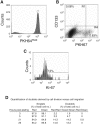
Tracking CD133+ cells in monolayer culture. (A) Nonselected primary epithelial cells were stained with PKH67, and a homogeneous subset of PI PKH67+ cells was isolated using a narrow band of fluorescence. (B) and (C) Cells isolated above were plated onto collagen 1 dishes with stem cell media (SCM) and irradiated STO feeders. After 4 days, the PKH67 cells were stained with anti-CD133-APC (B) and anti-Ki-67 (C). C is the R1 region (CD133+/PKH67) from B. Cells cultured in the presence of Colcemid® were used to establish the range of fluorescence exhibited by cells that had not divided. Experiments were carried out on 3 patients’ samples. One representative experiment is shown. (D) PKH26 (red)-labeled and PKH67 (green) CD133+ cells were mixed together in a 1:1 ratio and then plated, as above. At 3–26 h, the cells were mapped and red and green cells were counted. To calculate the number of doublets arising from cells division compared to cell migration/aggregation, we counted the number of 2 red cells, 2 green cells, and the number of 1 red and 1 green doublet.

Similar articles

Cited by

References

    1. Fuchs E, Tumbar T, Guasch G. Socialising with the Neighbors: stem cells and their niche. Cell. 2006;116:769–778. - PubMed
    1. Collins AT, Berry PA, Hyde C, Stower MJ, Maitland NJ. Prospective identification of tumorigenic prostate cancer stem cells. Cancer Res. 2005;65:10946–10951. - PubMed
    1. Birnie R, Bryce SD, Roome C, Dussupt V, Droop A, Lang SH, Berry PA, Hyde CF, Lewis JL, Stower MJ, Maitland MJ, Collins AC. Gene expression profiling of human prostate cancer stem cells reveals a pro-inflammatory phenotype and the importance of extracellular matrix interactions. Genome Biol. 2008;9:R83. - PMC - PubMed
    1. Collins AT, Habib FK, Maitland NJ, Neal DE. Identification and isolation of human prostate epithelial stem cells based on alpha(2)beta(1)-integrin expression. J Cell Sci. 2001;114(Pt 21):3865–3872. - PubMed
    1. Richardson GD, Robson CN, Lang SH, Neal DE, Maitland NJ, Collins AT. CD133, a novel marker for human prostatic epithelial stem cells. J Cell Sci. 2004;117(Pt 16):3539–3545. - PubMed

Publication types

MeSH terms