

Evaluation of localized and systemic immune responses in cutaneous leishmaniasis caused by Leishmania tropica: interleukin-8, monocyte chemotactic protein-1 and nitric oxide are major regulatory factors
- PMID: 20102417
- PMCID: PMC2878464
- DOI: 10.1111/j.1365-2567.2009.03223.x
Evaluation of localized and systemic immune responses in cutaneous leishmaniasis caused by Leishmania tropica: interleukin-8, monocyte chemotactic protein-1 and nitric oxide are major regulatory factors
Abstract
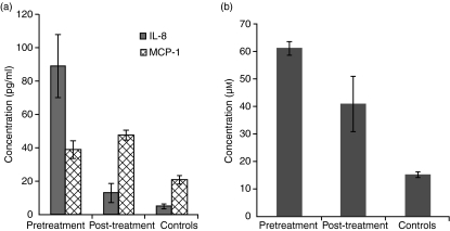
We have established Leishmania tropica as the causative agent of cutaneous leishmaniasis (CL) in the region of India where the disease is endemic. The association between localized and circulating levels of immune-determinants in CL patients was evaluated. Reverse transcription-polymerase chain reaction analysis revealed up-regulation of interferon-gamma (IFN-gamma), interleukin (IL)-1beta, IL-8, tumour necrosis factor-alpha (TNF-alpha), IL-10 and IL-4 in dermal lesions at the pretreatment stage (n = 31) compared with healthy controls (P < 0.001) and a significant down-regulation after treatment (n = 14, P < 0.05). The results indicated that an unfavourable clinical outcome in CL was not related to an inadequate T helper 1 (Th1) cell response, but rather to impairment in multiple immune functions. Comparative assessment of treatment regimes with rifampicin (RFM) or sodium antimony gluconate (SAG) revealed tissue cytokine levels to be significantly reduced after treatment with RFM (P < 0.005), while no significant decrease was evident in the levels of IFN-gamma, TNF-alpha and IL-10 (P > 0.05) as a result of treatment with SAG. Increased transcripts of monocyte chemoattractant protein-1 (MCP-1) (P < 0.001) and inducible nitric oxide synthase (iNOS) (P < 0.05) were evident before treatment in tissue lesions and remained high after treatment. Immunohistochemistry demonstrated strong expression of myeloperoxidase (MPO) and IL-8, and moderate expression of iNOS in dermal lesions. The expression levels of IL-8, MCP-1 and nitric oxide (NO) were high in patient sera before treatment, as determined using cytokine bead array and enzyme-linked immunosorbent assay (ELISA). At the post-treatment stage, the serum IL-8 levels had decreased; however, the levels of MCP-1 and NO remained high. These data suggest that IL-8 is an effector immune-determinant in the progression of CL, whereas NO facilitates the parasite killing by macrophages via MCP-1-mediated stimulation.
Figures


References
-
- Gramiccia M, Gradoni L. The current status of zoonotic leishmaniasis and approaches to disease control. Int J Parasitol. 2005;35:1169–80. - PubMed
-
- Schwenkenbecher JM, Wirth T, Schnur LF, et al. Microsatellite analysis reveals genetic structure of Leishmania tropica. Int J Parasitol. 2006;36:237–46. - PubMed
-
- Kumar R, Bumb RA, Ansari NA, Mehta RD, Salotra P. Cutaneous leishmaniasis caused by Leishmania tropica in Bikaner, India: parasite identification and characterization using molecular and immunological tools. Am J Trop Med Hyg. 2007;76:896–901. - PubMed
-
- Louis J, Himmelrich H, Parra-Lopez C, Tacchini-Cottier F, Launois P. Regulation of protective immunity against Leishmania major in mice. Curr Opin Immunol. 1998;10:459–64. - PubMed
Publication types
MeSH terms
Substances
LinkOut - more resources
Full Text Sources
Research Materials
Miscellaneous
