. 2010 Jan 26:11:11.
doi: 10.1186/1471-2199-11-11.

Correlation between ribonucleoside-diphosphate reductase and three replication proteins in Escherichia coli

Affiliations

Correlation between ribonucleoside-diphosphate reductase and three replication proteins in Escherichia coli

M Antonia Sánchez-Romero et al. BMC Mol Biol. .

Abstract

Background: There has long been evidence supporting the idea that RNR and the dNTP-synthesizing complex must be closely linked to the replication complex or replisome. We contributed to this body of evidence in proposing the hypothesis of the replication hyperstructure. A recently published work called this postulate into question, reporting that NrdB is evenly distributed throughout the cytoplasm. Consequently we were interested in the localization of RNR protein and its relationship with other replication proteins.

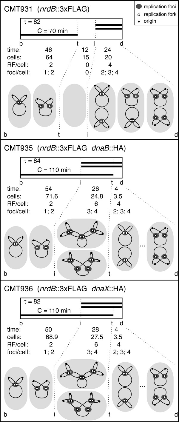
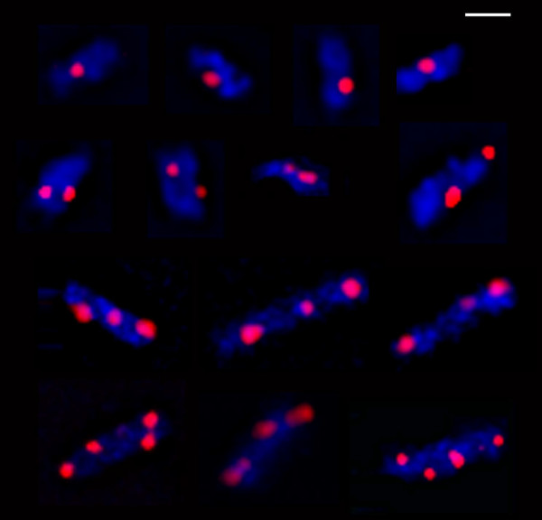
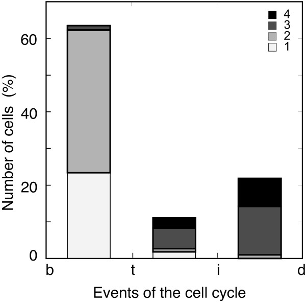
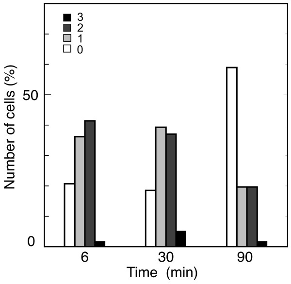
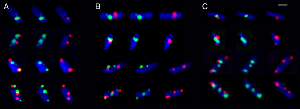
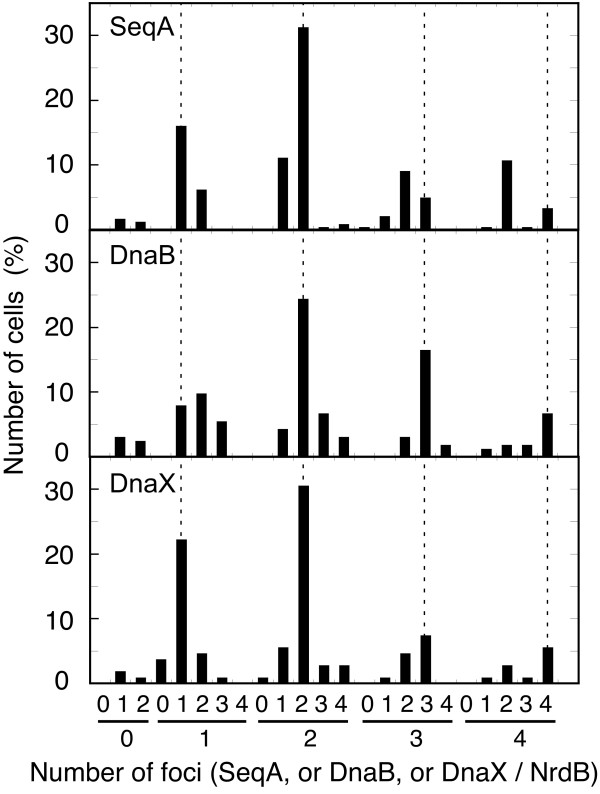
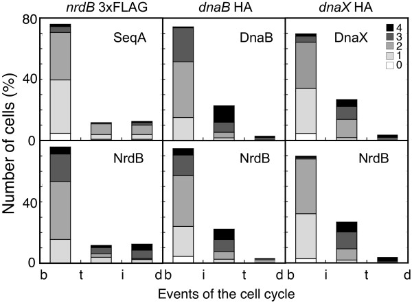
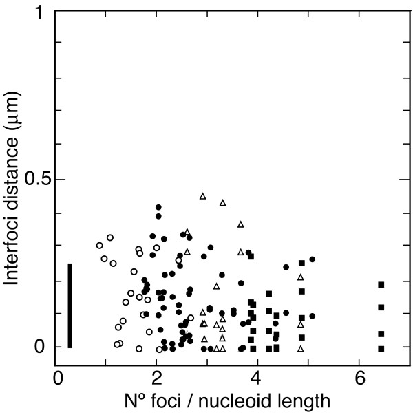
Results: We tagged NrdB protein with 3xFLAG epitope and detected its subcellular location by immunofluorescence microscopy. We found that this protein is located in nucleoid-associated clusters, that the number of foci correlates with the number of replication forks at any cell age, and that after the replication process ends the number of cells containing NrdB foci decreases.We show that the number of NrdB foci is very similar to the number of SeqA, DnaB, and DnaX foci, both in the whole culture and in different cell cycle periods. In addition, interfoci distances between NrdB and three replication proteins are similar to the distances between two replication protein foci.

Conclusions: NrdB is present in nucleoid-associated clusters during the replication period. These clusters disappear after replication ends. The number of these clusters is closely related to the number of replication forks and the number of three replication protein clusters in any cell cycle period. Therefore we conclude that NrdB protein, and most likely RNR protein, is closely linked to the replication proteins or replisome at the replication fork. These results clearly support the replication hyperstructure model.


Figures

Figure 1
Figure 1
Cell cycle events and chromosome structure. Cell cycle events and chromosome shapes relative to cell age in strains CMT931, CMT935, and CMT936 growing in glycerol minimal medium at 37°C. τ is the doubling time in minutes. C is the time in minutes required to replicate the chromosome. Thick bars represent replicating chromosomes. Time refers to the length in minutes of each of the three periods determined by cell birth (b), initiation of replication (i), termination of replication (t), and cell division (d). cells is the frequency of cells, in percentages, in each of the three periods. RF/cell is the number of replication forks per cell, and foci/cell is the expected number of foci per cell of a fluorescent replication protein depending on the association state of the sister replication forks, in each of the three cell cycle periods. The inset shows the symbols for the origin of replication (small black circle), for each replication fork (open circle), and for the foci of an immunostained replication protein (grey oval).
Figure 2
Figure 2
Fluorescence microscopy of tagged-NrdB cells. Fluorescence microscopy images of cells of CMT931 (nrdB::3×FLAG) immunolabeled with Cy3 conjugated anti-FLAG antibody (red) and stained with Hoechst 33258 (blue). The bar represents 1 μm.
Figure 3
Figure 3
NrdB foci and replication forks. Percentage of cells containing different numbers of NrdB::3×FLAG foci per cell of strain CMT931, distributed into the three periods of the cell cycle defined as from birth (b) to termination of replication (t), from termination to initiation (i), and from initiation to cell division (d), as described in Figure 1. Number of cells analyzed was 426. The inset shows the colour code for the four numbers of foci per cell.
Figure 4
Figure 4
Analysis of NrdB foci during synchronous replication. Number of NrdB foci in a CMT934 (nrdB::×FLAG dnaC2) culture after the three-step temperature shifts for synchronous initiation of one round of chromosome replication. Samples were withdrawn at 6, 30, and 90 min after the beginning of the 6 min period at 30°C to allow one initiation round. Number of cells analyzed was 261. The inset shows the colour code for the number of foci per cell.
Figure 5
Figure 5
Fluorescence microscopy of tagged-NrdB and SeqA, or DnaB, or DnaX cells. Fluorescence microscopy images of cells of CMT935 (nrdB::3×FLAG dnaB::HA) (A) or CMT936 (nrdB::3×FLAG dnaX::HA) (B) immunolabeled with Cy3-anti-FLAG (red) and FITC-anti-HA (green), or CMT931 (nrdB::3×FLAG) immunolabeled with Cy3 conjugated anti-FLAG (red) and FITC-anti-SeqA (green) antibodies (C). Cells were also stained with Hoechst 33258 for nucleoid visualization. Each group of cells shows nucleoid (blue) and, from left to right: both green and red, only green, and only red fluorescence. The bar represents 1 μm.
Figure 6
Figure 6
NrdB foci related to SeqA, DnaB, and DnaX foci. Frequency of the number of foci of NrdB together with SeqA fluorescent proteins in strain CMT931 (nrdB::3×FLAG), or with DnaB helicase in strain CMT935 (nrdB::3×FLAG dnaB::HA), or with DNA polymerase III τ subunit in strain CMT936 (nrdB::3×FLAG dnaX::HA) in mid log cells growing in glycerol minimal medium at 37°C. Numbers in the upper position of the horizontal axis are the number of foci of any of the three replication proteins present in a cell containing the number of NrdB foci indicated by the number in the lower position. Dotted lines identify cells with the same number of both foci. Numbers of cells analyzed were 129, 172, and 110, respectively.
Figure 7
Figure 7
NrdB foci and replication proteins foci along the cell cycle. Percentage of cells containing different numbers of foci during the three cell cycle periods described in Figure 1. Foci of fluorescent proteins were from SeqA and NrdB in strain CMT931 (nrdB::3×FLAG); DnaB helicase and NrdB in strain CMT935 (nrdB::3×FLAG dnaB::HA); and DnaX τ subunit and NrdB in strain CMT936 (nrdB::3×FLAG dnaX::HA). Numbers of cells analyzed were 129, 172, and 110, respectively. The inset shows the colour code for the number of foci per cell.
Figure 8
Figure 8
Distances between NrdB and DnaX foci relative to foci per cell. Interfoci distances between NrdB and DnaX τ subunit relative to foci compaction in cells containing 1 (open circles), 2 (solid circles), 3 (open triangles), or 4 (squares) pairs of foci. Foci compaction is expressed by the number of pairs of foci per nucleoid length. Vertical bar represents the focus diameter. Number of cells analyzed was 110.

Similar articles

Cited by

References

    1. Marians KJ. Understanding how the replisome works. Nat Struct Mol Biol. 2008;15:125–127. doi: 10.1038/nsmb0208-125. - DOI - PubMed
    1. Eklund H, Uhlin U, Färnegardh M, Logan DT, Nordlund P. Structure and function of the radical enzyme ribonucleotide reductase. Prog Biophys Mol Biol. 2001;77:177–268. doi: 10.1016/S0079-6107(01)00014-1. - DOI - PubMed
    1. Nordlund P, Reichard P. Ribonucleotide reductases. Annu Rev Biochem. 2006;75:681–706. doi: 10.1146/annurev.biochem.75.103004.142443. - DOI - PubMed
    1. Mathews CK, Sinha NK. Are DNA precursors concentrated at replication sites? Proc Natl Acad Sci USA. 1982;79:302–306. doi: 10.1073/pnas.79.2.302. - DOI - PMC - PubMed
    1. Warner HR. Properties of ribonucleoside disphosphate reductase in nucleotide-permeable cells. J Bacteriol. 1973;115:18–22. - PMC - PubMed

Publication types

MeSH terms

LinkOut - more resources