. 2010;12(1):R14.
doi: 10.1186/ar2915. Epub 2010 Jan 26.

CD16 (FcRgammaIII) as a potential marker of osteoclast precursors in psoriatic arthritis

Affiliations

CD16 (FcRgammaIII) as a potential marker of osteoclast precursors in psoriatic arthritis

Yahui Grace Chiu et al. Arthritis Res Ther. 2010.

Abstract

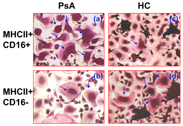
Introduction: Psoriatic arthritis (PsA) is a chronic inflammatory arthritis characterized by bone erosion mediated by osteoclasts (OC). Our previous studies showed an elevated frequency of OC precursors (OCP) in PsA patients. Here, we examined if OC arise from CD16-positive monocytes in PsA.

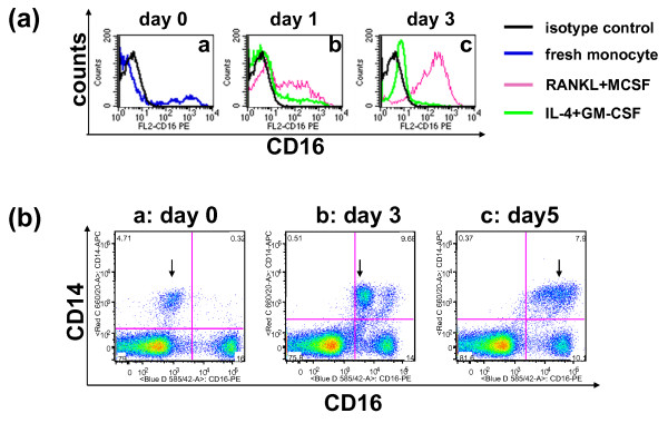
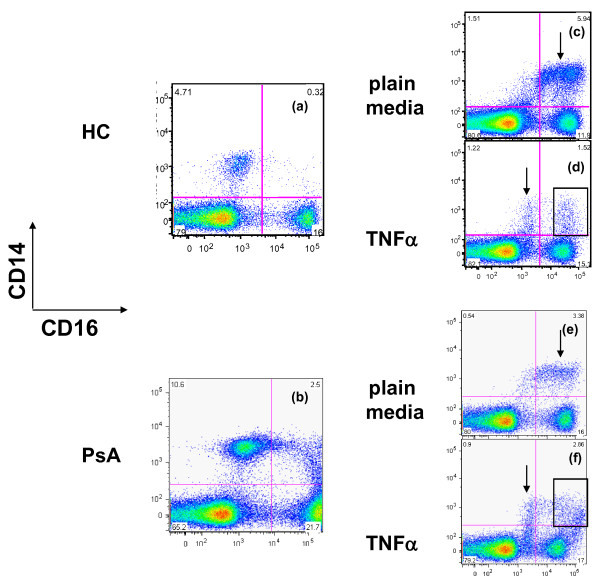
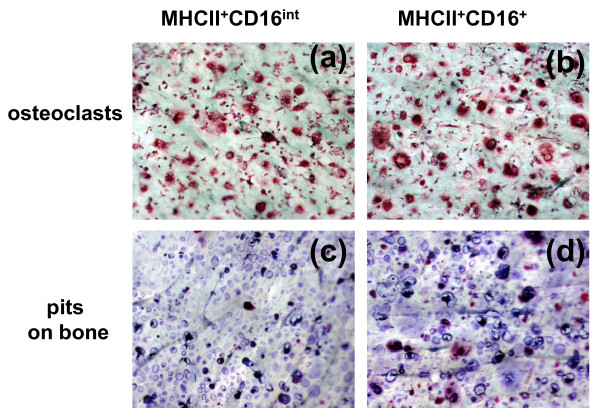
Methods: Peripheral blood mononuclear cells (PBMC) or monocytes were isolated from human peripheral blood and sorted based on CD16 expression. Sorted cells were cultured alone or with bone wafers in the presence of receptor activator of nuclear factor kappa-B ligand (RANKL) and macrophage colony-stimulating factor (M-CSF). Enumeration and bone erosion activity of OC were examined after culture. The effects of tumor necrosis factor-alpha (TNFalpha), OC-promoting (M-CSF plus RANKL), and dendritic cell (DC)-promoting (GM-CSF plus interleukin (IL)-4) cytokines on CD16 surface expression were examined by flow cytometry.

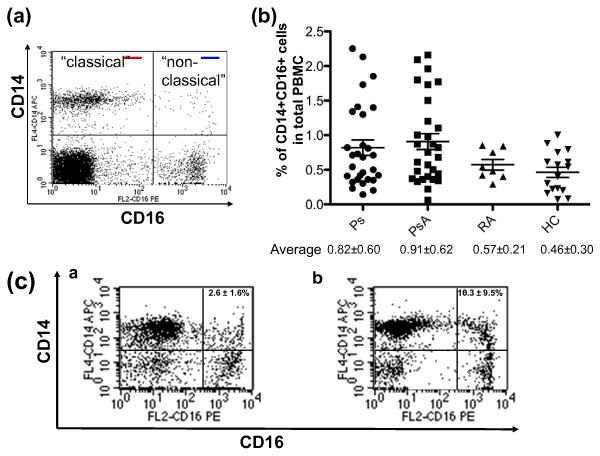
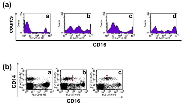
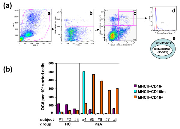
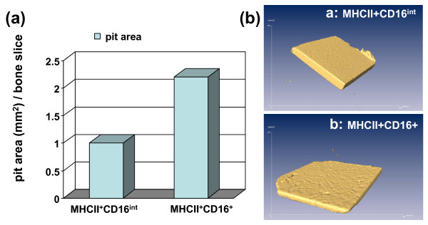
Results: PsA and psoriasis (Ps) subjects had a higher percentage of circulating inflammatory CD14+CD16+ cells than healthy controls (HC). Exposure of cells to OC-promoting, but not DC-promoting media, was associated with CD16 up-regulation. PBMC of Ps and PsA had a higher frequency of cells expressing intermediate levels of CD16. OC were mainly derived from CD16+ cells in PsA. Increased CD16 expression was associated with a higher bone erosion activity in PsA.

Conclusions: An increased frequency of circulating CD14+CD16+ cells was noted in PsA compared to controls, and intermediate levels of CD16 may suggest a transitional state of OCP during osteoclastogenesis. Intriguingly, TNFalpha blocked CD16 expression on a subset of CD14+ monocytes. Collectively, our data suggest that CD16 has the potential to serve as an OCP marker in inflammatory arthritis.


Figures

Figure 1
Figure 1
Ps and PsA patients have a higher percentage of CD14+CD16+ cells. Human peripheral blood mononuclear cells (PBMC) were isolated from peripheral blood, stained with an antibody cocktail composed of CD14-APC, CD16-PE, 7AAD, and analyzed by flow cytometry. Dead cells were excluded by 7AAD+. (a) Classical (CD14+CD16-, red line) and non-classical (CD14+CD16+, blue line) monocytes were labeled based on the classification by Strauss-Ayali and colleagues [5]. (b) The percentage of CD14+CD16+ cells in the PBMC of 16 healthy controls (HC), and 29 psoriasis (Ps), 28 psoriatic arthritis (PsA), and 8 rheumatoid arthritis (RA) patients. (c) The percentage of CD14+CD16+ cells in enriched human monocytes from HC and PsA. Monocytes were enriched by the Human Monocyte Enrichment Cocktail. The percentage of CD14+CD16+ cells in enriched monocytes from HC (2.6 ± 1.6%) and PsA patients (10.3 ± 9.5%) are shown in [a] and [b], respectively. The data are representative of 10 independent experiments.
Figure 2
Figure 2
Cytokines alter the cell surface expression of CD16 and human CD14+ cells undergo a transitional stage of CD16 up-regulation in OC-promoting culture conditions. (a) Enriched human monocytes were cultured in osteoclast (OC)-promoting media (receptor activator of nuclear factor kappa-B ligand (RANKL) + macrophage colony-stimulating factor (M-CSF), pink line) or dendritic cell (DC)-promoting media (IL-4 + granulocyte-macrophage colony-stimulating factor (GM-CSF), green line). Freshly isolated monocytes (blue line) and the isotype control (black line) are also shown. Surface expression of CD16 was monitored by FACS analysis on [a] day 0, [b] day 1, and [c] day 3, respectively. (b) Human peripheral blood mononuclear cells (PBMC) were cultured in OC-promoting media (RANKL + M-CSF) and the cell surface expression of CD14 and CD16 was monitored on [a] day 0, [b] day 3, and [c] day 5. Data shown here are live cells after forward scatter/side scatter (FSC/SSC) gating followed by dead cell exclusion using 7-amino-actinomycin D (AAD). Numbers shown in each quadrant are the percentage of total gated cells.
Figure 3
Figure 3
Cells that express intermediate levels of CD16 (CD16int) are found in vivo in human PBMC. (a) CD16 expression on human peripheral blood mononuclear cells (PBMC) is heterogeneous. The data shown here are the representatives of CD16 expression patterns on fresh human PBMC from 82 subjects. [a] PBMC that expressed high and low levels of CD16 without the CD16int population. [b to d] PBMC that have CD16int populations with different CD16 expression levels. (b) Ex vivo analysis of CD14 and CD16 expression. Representative examples of individuals [a] without a CD16int population, [b] CD14- CD16int (arrow) cells and [c] CD14+ CD16int (arrow) cells are shown. In total, 16 healthy control (HC), 29 psoriasis (Ps), 29 psoriatic arthritis (PsA), and 8 rheumatoid arthritis (RA)patients were included in this analysis. See Table 1 for the summary of CD16int percentage in HC, Ps, PsA and RA groups.
Figure 4
Figure 4
CD16- and CD16+ cells are the major reservoirs of OC precursors in HC and PsA patients, respectively. (a) 30 to 50% of MHCII+CD16+ cells are CD14+. Human peripheral blood mononuclear cells (PBMC) were isolated from the peripheral blood and stained with an antibody cocktail composed of MHCII-FITC, CD14-APC, CD16-PE, 7AAD. The gating strategy is sequentially illustrated from a to d. [a] PBMC were gated by FSC/SSC; [b] live cells were gated by 7AAD; [c] dot plot analysis of MHCII and CD16 expression on live cells; [d] the expression of CD14 on MHCII+CD16+ cells gated from c. Red line is the isotype control and blue line is staining from CD14-APC. The number shown in the histogram represents the frequency of CD14+ cells per total MHCII+CD16+ cells. [e] The relative percentage of CD14+CD16+ to MHCII+CD16+ cells. Sample shown here is the representative of 3 healthy controls (HC). (b) Human PBMC from HC and psoriatic arthritis (PsA) patients were isolated, stained with antibodies, and sterile sorted into three populations, MHCII+CD16-, MHCII+CD16int, and MHCII+CD16+. Osteoclastogenesis was examined by osteoclast (OC) count after eight-day culture in the presence of receptor activator of nuclear factor kappa-B ligand (RANKL) and macrophage colony-stimulating factor (M-CSF) (MHCII+CD16-: purple, MHCII+CD16int: turquoise, MHCII+CD16+: orange). Subject numbers 1 to 3 are HC and numbers 4 to 8 are PsA patients.
Figure 5
Figure 5
OC derived from MHCII+CD16+ and MHCII+CD16- cells of HC and PsA patients have distinct phenotypes. Peripheral blood mononuclear cells (PBMC) isolated from (a and b) psoriatic arthritis (PsA) and (c and d) healthy controls (HC) and were sterile sorted into (a and c) MHCII+CD16+ and (b and d) MHCII+CD16- populations. Sorted cells were cultured in the presence of receptor activator of nuclear factor kappa-B ligand (RANKL) and macrophage colony-stimulating factor (M-CSF) for eight days, and tartrate-resistant acid phosphatase stained for osteoclast (OC) quantification. Arrows indicate mature OC. The details of differences in OC generation potential and numbers of nuclei from these sorted subsets are summarized in Table 4.
Figure 6
Figure 6
CD14+cells from HC and PsA respond differently to TNFα in regards to CD16 cell surface expression. The expression of CD16 on freshly isolated peripheral blood mononuclear cells (PBMC) from healthy control (HC) and psoriatic arthritis (PsA) was shown in (a) and (b), respectively. After PBMC were cultured in the (c and e) absence or (d and f) presence of TNFα for three days, cell surface expression of CD16 was examined again using the same antibody cocktail composed of anti-CD16 (PE), anti-CD14 (APC), anti-CD3 (Pacific Blue), and anti-CD19 (APC-Cy7) antibodies with 7AAD. The anti-CD3, anti-CD19 antibodies and 7AAD were included to exclude T cells, B cells, and dead cells, respectively. Numbers shown in each quadrant (c to f) represent the percentage of cells per total gated live cells. Data shown here are the representative of two HC and three PsA samples.
Figure 7
Figure 7
Elevated CD16 surface expression was associated with increased bone erosion activity in PsA monocytes. Sterile sorted MHCII+CD16+ and MHCII+CD16int cells were co-cultured with bone slices in the presence of receptor activator of nuclear factor kappa-B ligand (RANKL) and macrophage colony-stimulating factor (M-CSF) for 14 days. (a and b) Osteoclast (OC) were identified by tartrate-resistant acid phosphatase (TRAP) staining, and (c and d) bone erosion pits were visualized after toluidine blue staining.
Figure 8
Figure 8
CD16+ cells have a higher bone resorption activity than CD16int cells. Sterile sorted MHCII+CD16+ and MHCII+CD16int cells were co-cultured with bone slices in the presence of receptor activator of nuclear factor kappa-B ligand (RANKL) and macrophage colony-stimulating factor (M-CSF) for 14 days. (a) Quantification of bone pits eroded by MHCII+CD16int and MHCII+CD16+ cells. (b) Reconstructed three-dimensional micro-CT images of the bone wafer incubated with MHCII+CD16int [a] and MHCII+CD16+ [b] sorted cells. The data are representative of three independent sorting experiments.

Similar articles

Cited by

References

    1. Kane D, Pathare S. Early psoriatic arthritis. Rheum Dis Clin North Am. 2005;31:641–657. doi: 10.1016/j.rdc.2005.07.009. - DOI - PubMed
    1. Teitelbaum SL, Ross FP. Genetic regulation of osteoclast development and function. Nat Rev Genet. 2003;4:638–649. doi: 10.1038/nrg1122. - DOI - PubMed
    1. Ritchlin CT, Haas-Smith SA, Li P, Hicks DG, Schwarz EM. Mechanisms of TNF-alpha- and RANKL-mediated osteoclastogenesis and bone resorption in psoriatic arthritis. J Clin Invest. 2003;111:821–831. - PMC - PubMed
    1. Ziegler-Heitbrock HW. Heterogeneity of human blood monocytes: the CD14+ CD16+ subpopulation. Immunol Today. 1996;17:424–428. doi: 10.1016/0167-5699(96)10029-3. - DOI - PubMed
    1. Strauss-Ayali D, Conrad SM, Mosser DM. Monocyte subpopulations and their differentiation patterns during infection. J Leukoc Biol. 2007;82:244–252. doi: 10.1189/jlb.0307191. - DOI - PubMed

Publication types