. 2010 May;29(4):261-75.
doi: 10.1016/j.matbio.2010.01.006. Epub 2010 Jan 25.

Characterization of the six zebrafish clade B fibrillar procollagen genes, with evidence for evolutionarily conserved alternative splicing within the pro-alpha1(V) C-propeptide

Affiliations

Characterization of the six zebrafish clade B fibrillar procollagen genes, with evidence for evolutionarily conserved alternative splicing within the pro-alpha1(V) C-propeptide

Guy G Hoffman et al. Matrix Biol. 2010 May.

Abstract

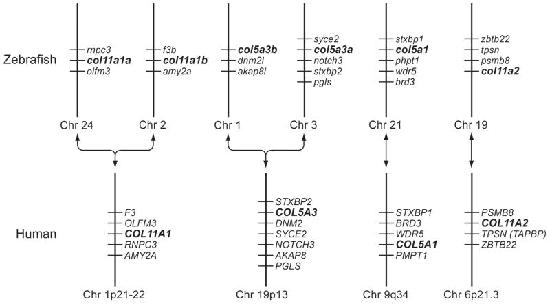
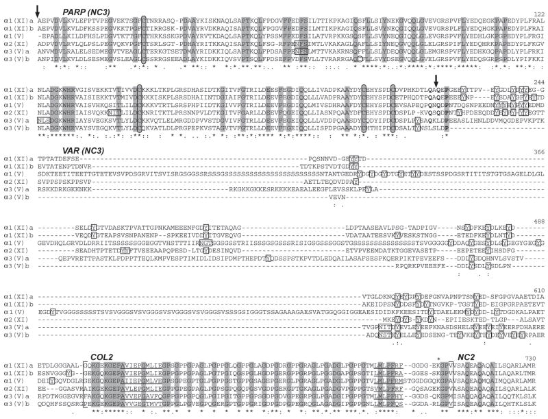
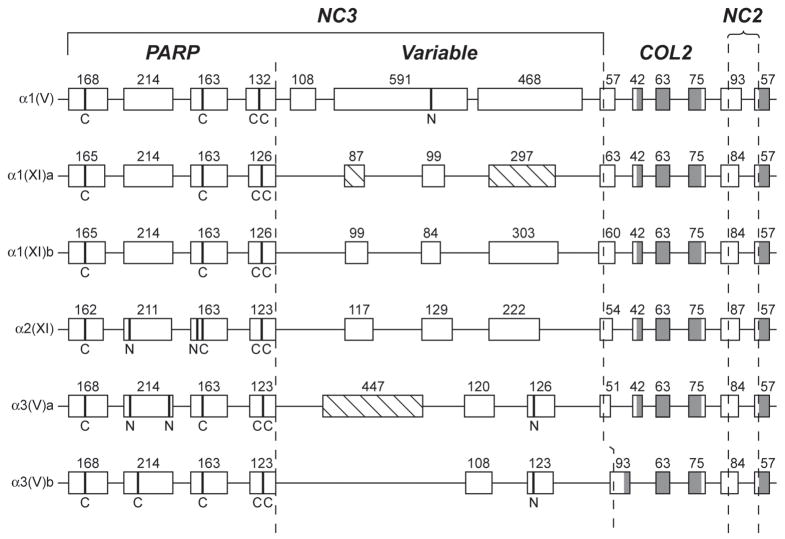
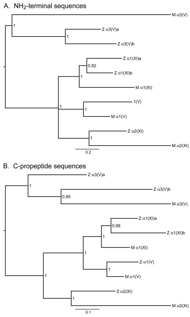
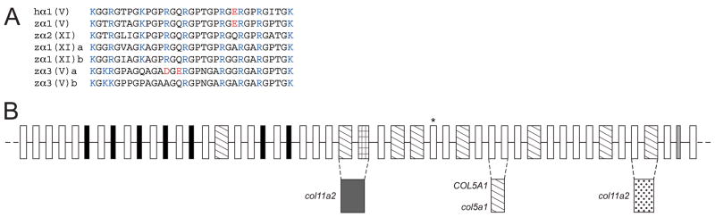
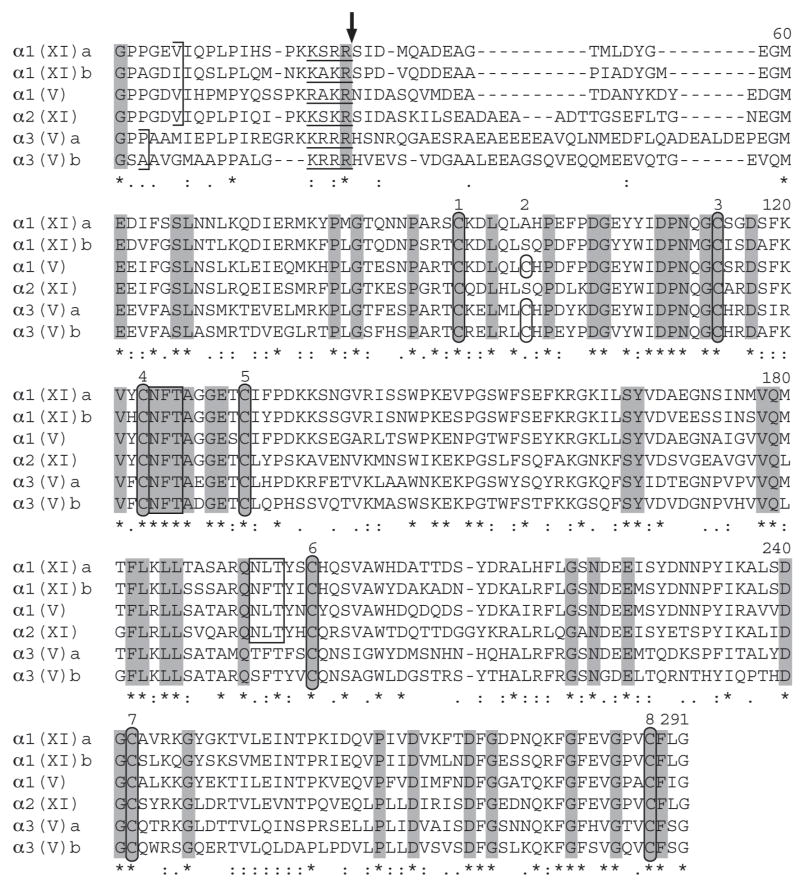
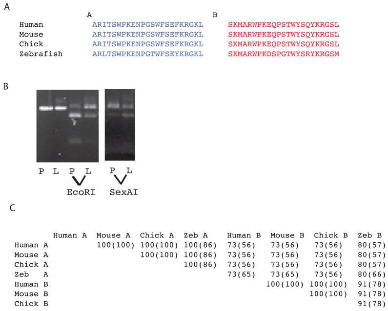
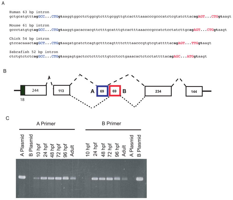
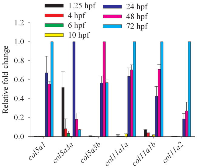
Genes for tetrapod fibrillar procollagen chains can be divided into two clades, A and B, based on sequence homologies and differences in protein domain and gene structures. Although the major fibrillar collagen types I-III comprise only clade A chains, the minor fibrillar collagen types V and XI comprise both clade A chains and the clade B chains pro-alpha1(V), pro-alpha3(V), pro-alpha1(XI) and pro-alpha2(XI), in which defects can underlie various genetic connective tissue disorders. Here we characterize the clade B procollagen chains of zebrafish. We demonstrate that in contrast to the four tetrapod clade B chains, zebrafish have six clade B chains, designated here as pro-alpha1(V), pro-alpha3(V)a and b, pro-alpha1(XI)a and b, and pro-alpha2(XI), based on synteny, sequence homologies, and features of protein domain and gene structures. Spatiotemporal expression patterns are described, as are conserved and non-conserved features that provide insights into the function and evolution of the clade B chain types. Such features include differential alternative splicing of NH(2)-terminal globular sequences and the first case of a non-triple helical imperfection in the COL1 domain of a clade B, or clade A, fibrillar procollagen chain. Evidence is also provided for previously unknown and evolutionarily conserved alternative splicing within the pro-alpha1(V) C-propeptide, which may affect selectivity of collagen type V/XI chain associations in species ranging from zebrafish to human. Data presented herein provide insights into the nature of clade B procollagen chains and should facilitate their study in the zebrafish model system.


Figures

Fig. 1
Fig. 1
Apparent syntenic relationships between zebrafish and human clade B collagen genes. Apparent conserved synteny is dispayed for human gene COL11A1 with zebrafish loci NM_001083844 and XM_677653, of the human gene COL11A2 with locus NM_001079992, of the human gene COL5A1 with XM_685787, and of the human gene COL5A3 with both XM_001921860 and XM_688785; leading to provisional designation of the zebrafish loci as col11a1a, col11a1b, col11a2, col5a1, col5a3a and col5a3b, respectively. Changes in the order of genes in some instances between human and zebrafish is presumably due to differential chromosomal rearrangements during evolution. To determine conservation of synteny between zebrafish and human clade B procollagen chain genes, upstream and downstream genes on respective chromosomes in the NCBI ENTREZ D. rerio Tubingen and H. sapiens genome projects were manually compared.
Fig. 2
Fig. 2
Alignment of NH2-terminal sequences of zebrafish clade B procollagen chains. NH2-terminal sequences were aligned using the EMBL-EBI ClustalW2 server. Dashes represent gaps introduced for optimal sequence alignment. Vertical arrows mark the approximate site of signal peptide cleavage and cleavage by BMP1-like proteinases, based on predicted and demonstrated sites in mammalian clade B chains (Gopalakrishnan et al., 2004; Greenspan et al., 1991; Imamura et al., 2000; Imamura et al., 1998; Unsold et al., 2002). PARP, and variable (VAR) subdomains of noncollagenous domain 3 (NC3) are labeled, as is collagenous domain 2 (COL2), and noncollagenous domain 2 (NC2). The extent of COL2 is marked by brackets. Noncollagenous interruptions in the COL2 domain are underlined. Cysteines are circled. Tyrosines between the PARP and COL2 domains are boxed, as are potential Asn-linked glycosylation sites. Residues found at the pro-α1(V) BMP1-cleavage site and conserved in zebrafish clade B chains are in boldface type. Asterisks and dots at the bottom of the alignment signify extent of similarity of aligned sequences, with asterisks denoting identity at a given position in all six zebrafish clade B chains. Residues identical in all six zebrafish and all reported mammalian clade B procollagen chains (Imamura et al., 2000) are shaded.
Fig. 3
Fig. 3
Comparison of the intron/exon organizations of NH2-terminal sequence-encoding regions of zebrafish clade B procollagen genes. Boxes represent exons. Numbers represent lengths in basepairs. Shaded and open regions of boxes represent triple-helix and non-triple helix coding sequences, respectively. Dashed lines demarcate PARP-, Variable-, COL2- and NC2-encoding regions. Hatched boxes represent alternatively spliced exons. Sequences encoding cysteines and potential Asn-linked glycosylation sites are marked by broad bands and the letters “C” and “N”, respectively.
Fig. 4
Fig. 4
Bayesian majority-rule consensus phylograms for the NH2-terminal and C-propeptide regions of zebrafish and mammalian clade B procollagen chains. These midpoint rooted trees provide estimates of phylogenetic relationships among the N-terminal globular (A) and C-propeptide (B) amino acid sequences of these chains, with branch lengths drawn in proportion to the average number of substitutions per site on a given branch (scale bar provided). Numbers represent posterior probability values. Z and M designate zebrafish and mammalian chains, respectively. Mammalian sequences used were from human pro-α3(V) and pro-α1(V), and murine pro-α1(XI) and pro-α2(XI) chains. Both trees are consistent with hypothesized relatedness of the various genes hypothesized in the text. Agreement between the NH2-terminal and C-propeptide trees is very high, with identical topologies except for one polytomy in the NH2-terminal tree and one discrepancy in the relationships among pro-α3(V)a, pro-α3(V)b, and mammalian pro-α3(V). The NH2-terminal tree but not the C-propeptide tree is consistent with the hypothesis that the zebrafish pro-α3(V)a and pro-α3(V)b paralogs trace back to the whole genome duplication event that occurred early in the radiation of ray-finned fish. Although the optimal C-propeptide tree would seem to contradict this likely hypothesis, the posterior probability of the C-propeptide tree is low enough (0.95) that the hypothesis supported by the NH2-terminal region tree remains plausible. Comparison of branch lengths with associated scale bars for the two trees indicates different rates of evolutionary change, consistent with differences in the intensity of purifying selection for NH2-terminal and C-propeptide sequences.
Fig. 5
Fig. 5
Features of the COL1-encoding regions of zebrafish clade B procollagen genes. (A) An alignment is shown of zebrafish clade B procollagen COL1 sequences corresponding to the human α1(V) heparin-binding domain. Basic and acidic residues are blue and red, respectively. (B) White, black, hatched, dark grey, light grey, checkered and stippled boxes represent 54-, 45-, 108-, 198-, 36-, 90-, and 162-bp exons, respectively. Dashed lines indicate fusion of 108- and 90-bp exons found in other clade B chain genes to form a 198-bp exon in zebrafish col11a2, fusion of two 54-bp exons found in other clade B chain genes to form a 108-bp exon in human COL5A1 and zebrafish col5a1, and fusion of 54- and 108-bp exons found in other clade B chain genes to form a 162-bp exon in zebrafish col11a2. An asterisk marks the exon encoding an imperfection of the triple helix in zebrafish col11a1a.
Fig. 6
Fig. 6
Alignment of C-propeptide sequences of zebrafish clade B procollagen chains. C-propeptide sequences were aligned using the EMBL-EBI ClustalW2 server. Dashes represent gaps introduced for optimal alignment. Ends of COL1 domains are marked by brackets. Consensus sites for cleavage by proprotein convertases are underlined, and a vertical arrow marks the predicted site for cleavage. Cysteines are circled and potential Asn-linked glycosylation sites are boxed. Asterisks and dots at the bottom of the alignment signify extent of similarity of aligned sequences, with asterisks denoting identity in all six zebrafish clade B chains. Residues identical in all six zebrafish and all reported mammalian clade B procollagen chains (Imamura et al., 2000) are shaded.
Fig. 7
Fig. 7
Alternative splicing in the COL5A1 C-propeptide region and conservation of alternatively spliced exon sequences across species. (A) Amino acid sequences are shown for exons A and B for human mouse, chick and zebrafish. (B) Restriction with EcoRI, which cuts within exon A, but not exon B sequences, or SexAI, which cuts within exon B but not exon A sequences, demonstrates varying ratios of exon A and B sequences in human placenta (P) and liver (L). The first two lanes are uncut cDNA. (C) Percentages of similarity and identity (parentheses) between the amino acid sequences of exons A and B in the pro-α1(V) genes of human, mouse, chick, and zebrafish were obtained by alignment using the BLAST Basic Local Alignment Search Tool.
Fig. 8
Fig. 8
(A) Mutually exclusive alternative splicing of exons A and B in pro-α1(V) genes of zebrafish and other species. Sequences are shown of splice junctions and introns separating exons A (blue) and B (red) in the C-propeptide regions of pro-α1(V) genes of human, mouse, chick, and zebrafish. Exonic sequences are uppercase and intronic sequences are lower case. Invariable 5′ gt and 3′ ag ends of introns are in boldface type. (B) A model is shown for the proposed mutually exclusive alternative splicing of exons A and B. (C) RT-PCR with exon A- and B-specific primers demonstrates expression of both exons in col5a1 transcripts of zebrafish embryos and adults. PCR of plasmids containing cloned zebrafish exon A and B sequences (A plasmid and B plasmid, respectively) demonstrates specificity of the primers.
Fig. 9
Fig. 9
Temporal distribution of clade B procollagen gene expression throughout embryogenesis. Real time quantitative RT-PCR analysis of RNA levels of all zebrafish clade B procollagen genes was performed for harvested embryos from 1.25 hpf (8-cell), 4 hpf (sphere), 6 hpf (shield), 10 hpf (bud), 24 hpf (prim-5), 48 hpf (long-pec), and 72 hpf (protruding mouth) stages. Levels of expression of each gene are given, relative to the stage at which they are maximally expressed, and normalized to expression of the β-actin housekeeping gene.
Fig. 10
Fig. 10
Spatial distribution of clade B procollagen gene expression in caudal regions of 30, 48 and 72 hpf embryos. (A) Whole mount in situ hybridization shows expression of col11a1b, col11a1a and col11a2, and col2a1 as a control, in the notochord of 30 (a-d), 48 (e and f), and 72 (g-i) hpf embryos. (B) Whole mount in situ hybridization shows expression of col5a1, col5a3a and col1a1 as a control, in the notochord of 30 (a-d), 48 (e and f) and 72 (g and h) hpf embryos. Note similar localization of col5a1 and col1a1 expression surrounding the notochord and between myotomes at 48 and 72 hpf (panels eh). Col1a1 (arrowhead), but not col5a1, expression, is strong in the caudal fin fold. A probe corresponding to Col5a3a exons 1-5 and 7 sequences detected expression at 30 hpf throughout the length of the notochord (panel b), whereas a probe corresponding to alternatively spliced Col5a3a exon 6 (splice) only detected expression in the caudal notochord (panel c) at this time.

Similar articles

Cited by

References

    1. Abedin MZ, Ayad S, Weiss JB. Isolation and native characterization of cysteine-rich collagens from bovine placental tissues and uterus and their relationship to types IV and V collagens. Biosci Rep. 1982;2:493–502. - PubMed
    1. Baas D, Malbouyres M, Haftek-Terreau Z, Le Guellec D, Ruggiero F. Craniofacial cartilage morphogenesis requires zebrafish col11a1 activity. Matrix Biol 2009 - PubMed
    1. Bernard M, Yoshioka H, Rodriguez E, Van der Rest M, Kimura T, Ninomiya Y, Olsen BR, Ramirez F. Cloning and sequencing of pro-alpha 1 (XI) collagen cDNA demonstrates that type XI belongs to the fibrillar class of collagens and reveals that the expression of the gene is not restricted to cartilagenous tissue. J Biol Chem. 1988;263:17159–66. - PubMed
    1. Birk DE, Fitch JM, Babiarz JP, Doane KJ, Linsenmayer TF. Collagen fibrillogenesis in vitro: interaction of types I and V collagen regulates fibril diameter. J Cell Sci. 1990;95(Pt 4):649–57. - PubMed
    1. Boot-Handford RP, Tuckwell DS. Fibrillar collagen: the key to vertebrate evolution? A tale of molecular incest. Bioessays. 2003;25:142–51. - PubMed

Publication types

MeSH terms

LinkOut - more resources