. 2010 Apr 2;285(14):10890-901.
doi: 10.1074/jbc.M109.058347. Epub 2010 Jan 26.

Xenopus skip modulates Wnt/beta-catenin signaling and functions in neural crest induction

Affiliations

Xenopus skip modulates Wnt/beta-catenin signaling and functions in neural crest induction

Ying Wang et al. J Biol Chem. .

Abstract

The beta-catenin-lymphoid enhancer factor (LEF) protein complex is the key mediator of canonical Wnt signaling and initiates target gene transcription upon ligand stimulation. In addition to beta-catenin and LEF themselves, many other proteins have been identified as necessary cofactors. Here we report that the evolutionally conserved splicing factor and transcriptional co-regulator, SKIP/SNW/NcoA62, forms a ternary complex with LEF1 and HDAC1 and mediates the repression of target genes. Loss-of-function studies showed that SKIP is obligatory for Wnt signaling-induced target gene transactivation, suggesting an important role of SKIP in the canonical Wnt signaling. Consistent with its involvement in beta-catenin signaling, the C-terminally truncated forms of SKIP are able to stabilize beta-catenin and enhance Wnt signaling. In Xenopus embryos, both overexpression and knockdown of Skip lead to reduced neural crest induction, consistent with down-regulated Wnt signaling in both cases. Our results indicate that SKIP is a novel component of the beta-catenin transcriptional complex.


Figures
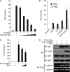
FIGURE 1.
FIGURE 1.
Skip inhibits Wnt/β-catenin signaling in HEK293T cells. A, full-length Skip inhibits β-catenin signaling in a dose-dependent manner. TOPflash reporter assays were carried out in HEK293T cells. β-Catenin and 0.1, 1, 2, 5, or 10 ng of skip plasmids were transfected as indicated. B, Wnt1- and LiCl-induced Wnt signaling are also reduced by co-transfection of Skip. 20 mm LiCl was used to treat cells for 12 h. C, Skip inhibits S37A-β-catenin signaling in a dose-dependent manner. 293T cells were transfected in each well with S37A-β-catenin and/or 5 or 10 ng of skip plasmids as indicated. D, neither mRNA nor protein of β-catenin was affected by overexpression of Skip. Skip, GFP, and β-catenin plasmids were transfected in 293T cells as indicated. 48 h later, cells were analyzed by Western blot (WB) and RT-PCR simultaneously. Actin and glyceraldehyde-3-phosphate dehydrogenase (GAPDH) were used as loading controls. IB, immunoblot.
FIGURE 2.
FIGURE 2.
Skip inhibits neural crest development. A, Xenopus skip expression at the indicated development stages was analyzed by RT-PCR. −RT, minus reverse transcription. Histone H4 was used for normalization. B, expression profile of skip in Xenopus embryos was shown by whole mount in situ hybridization. st, stage. C, Skip inhibits β-catenin signaling in a dose-dependent manner in Xenopus embryos. 4-Cell-stage embryos were injected equatorially with 250 pg of β-catenin and/or 50, 250, or 500 pg of skip mRNA, together with a TOPflash reporter plasmid, in each blastomere as indicated. Embryos were gathered at stage 11.5 and analyzed by the Dual-Luciferase assay system. D, skip mRNA injection causes inhibition of slug expression. 2-Cell-stage embryos were injected in one blasteomere with control (Con) or skip mRNA (2 ng) and fixed at the early neurula stage for whole mount in situ hybridization analysis with slug, Sox3, and engrailed2 (en2) probes. lacZ mRNA (0.5 ng) was co-injected as a lineage tracer. Light blue, lacZ staining; dark blue and brown, hybridization signal. E, Skip blocks slug induction by Wnt signaling in Xenopus animal caps. 100 pg of noggin, 50 pg of wnt8, and 1 ng of skip mRNA were injected animally into each blastomere at the 4-cell-stage as indicated. At stage 8, animal caps were dissected, cultured until stage 14, and analyzed for expressions of slug, NCAM, Xbra, and H4 by RT-PCR. H4 was used as the loading control, and Xbra was used to confirm that the animal caps were without mesoderm contamination. −RT, minus reverse transcription; WE, whole embryo.
FIGURE 3.
FIGURE 3.
Skip forms a ternary complex with LEF1 and HDAC1. A, Skip interacts with TCF/LEF family members. HEK293T cells were transfected as indicated, and lysates were immunoprecipitated by FLAG-M2 beads. Total lysates and IP samples were analyzed by Western blot (IB, immunoblot). HC, heavy chain of immunoglobulin. B, Skip interacts with HDAC1. 293T cells were transfected as indicated, and IP was performed as in A. C, Skip, HDAC1, and LEF1 are in the same complex. 293T cells were transfected as indicated, and two-step IP was performed as described under “Experimental Procedures.” Equal amount of myc-xLef1 was recovered after the first-step IP (IP1); however, after the second-step IP (IP2), LEF1 was recovered only when all three proteins were co-transfected but not when HDAC1 was absent. D, Skip enhances the interaction between LEF1 and HDAC1. 293T cells were transfected as indicated, and IPs were performed with anti-Myc antibody. E, overexpressed Skip protein associates with the promoter of Wnt target gene. 293T cells were transfected as indicated, and ChIP was performed with the indicated antibodies followed by PCR with specific primers against WRE on the c-myc promoter.
FIGURE 4.
FIGURE 4.
Skip is required for Wnt signaling in HEK293T cells. A, two independent siRNAs against human SKIP efficiently down-regulate endogenous SKIP in 293T cells. 100 nm siRNAs were transfected, and 72 h later total cell lysates were examined by Western blot (IB, immunoblot) with antibody against endogenous human SKIP. B and C, SKIP siRNAs down-regulate both β-catenin and Wnt signaling. TOPflash signaling was tested 72 h after siRNA transfection. Con, control siRNA. D, Xenopus Skip partially rescues Wnt1 signaling reduced by SKIP siRNA in 293T cells. E, SKIP siRNAs do not affect Wnt3A conditioned medium-induced accumulation of β-catenin. Cytosolic fractions were subjected to Western blot. Wnt3A CM, Wnt3A conditioned medium. F, SKIP is required for Wnt signal-induced target gene expression in 293T cells. 293T cells were transfected with SKIP siRNAs and 48 h later were treated with LiCl for 12 h. AXIN2 expression was analyzed by real-time PCR.
FIGURE 5.
FIGURE 5.
Skip is required for neural crest induction in Xenopus embryos. A, antisense morpholino oligos against each of the two pseudoalleles of the X. laevis skip gene efficiently inhibit Skip translation. 4-Cell-stage embryos were injected equatorially with 2 ng of 5′-UTR1 mRNA, 1 ng of 5′-UTR2 mRNA, and 12.5 ng of MO in each blastomere as indicated. Embryos were cultured until stage 11.5 and analyzed by Western blot (IB, immunoblot). B, skip-MO injection causes failure of neural crest formation. 4-Cell-stage embryos were injected animally with 2.5 ng of MO and/or 1 ng of skip mRNA in each blastomere as indicated. C, the percentage of embryos with the phenotypes indicated in B is shown as bars (n > 40). D, skip-MO injection inhibits neural crest marker gene expression. Whole mount in situ hybridization analysis of slug, Sox3, and engrailed2 (en2) is shown. lacZ staining was used as a lineage tracer. The blue color stands for lacZ staining and the brown color for the hybridization signal. 2.5 ng of MO and/or 0.5 ng of lacZ mRNA was injected into one blastomere at the 2-cell stage. Early neurula stage embryos were shown in a dorsal view with the anterior side up. E, skip-MO injection blocks slug induction by Wnt signaling in animal caps. 100 pg of noggin, 50 pg of wnt8, and 2.5 and/or 5 ng of MO were injected animally into each blastomere at the 4-cell stage as indicated. At stage 8, animal caps were dissected, cultured until stage 14, and analyzed for expression of slug, NCAM, and H4 by RT-PCR. H4 was used as the loading control. −RT, minus reverse transcription; WE, whole embryo; Std, standard.
FIGURE 6.
FIGURE 6.
Domain analysis of Skip effect on Wnt signaling. A, schematic showing Skip deletions. B, cellular localization of Skip deletions. EGFP-tagged Skip and its deletions were transfected into HeLa cells. The nucleus was stained with 4,6-diamidino-2-phenylindole. C, Skip and its deletions affect Wnt signaling. The skip plasmids were co-transfected with vector, β-catenin, or Wnt as indicated, and a luciferase reporter assay was performed. D, the SNW domain likely mediates the interaction between Skip and LEF1. E, both the N-terminal and SNW domains are required for the interaction between Skip and HDAC1. Transfected 293T cells were lysed, immunoprecipitated by FLAG-M2 beads, and analyzed by Western blot (IB, immunoblot). HC, high chain of immunoglobulin; LC, light chain of immunoglobulin.
FIGURE 7.
FIGURE 7.
C-terminally truncated Skip enhances Wnt signaling. A, some of the Skip deletions cause β-catenin accumulation. Myc-β-catenin was co-transfected with Skip deletions in 293T cells, and cell lysates were analyzed by Western blot (IB, immunoblot). B, Skip-(1–341) promotes β-catenin accumulation without affecting its mRNA level or co-transfected GFP. Skip-(1–341), GFP, and β-catenin were transfected in 293T cells as indicated. Total cell suspension was divided into two parts; one was analyzed by Western blot (WB) and the other by RT-PCR. Actin and glyceraldehyde-3-phosphate dehydrogenase (GAPDH) were used as loading controls. C, β-catenin degradation is drastically blocked in the presence of Skip-(1–341). 100 μg/ml cycloheximide (CHX) was added 40 h after transfection; cells were then collected every 2 h, and the total lysates were analyzed by Western blot. D, Skip-(1–341) induces posteriorization of Xenopus embryos. 4-Cell-stage embryos were injected animally with 250 pg of skip-(1–341) mRNA and/or 10 pg of Wnt8 plasmid DNA in each blastomere as indicated. Tail bud-stage embryos were shown from a lateral view with the anterior side toward the left. A closer look at the head region is shown on the right. ppl, preprolactin was used as the injection control.
FIGURE 8.
FIGURE 8.
Skip colocalizes and weakly interacts with β-catenin. A, β-catenin is co-immunoprecipitated with Skip and its deletions. 293T cells was transfected as indicated, and total cell lysates were immunoprecipitated by anti-Myc antibody and analyzed by Western blot (IB, immunoblot). HC, heavy chain of immunoglobulin; LC, light chain of immunoglobulin, B, full-length Skip colocalizes with β-catenin in the nucleus. HeLa cells were co-transfected with EGFP-Skip and red fluorescent protein-β-catenin. The green color stands for Skip and red for β-catenin. The nucleus stained by 4,6-diamidino-2-phenylindole (DAPI) is shown in blue.

Similar articles

Cited by

References

    1. Logan C. Y., Nusse R. (2004) Annu. Rev. Cell Dev. Biol. 20, 781–810 - PubMed
    1. Moon R. T., Kohn A. D., De Ferrari G. V., Kaykas A. (2004) Nat. Rev. Genet. 5, 689–699 - PubMed
    1. Clevers H. (2006) Cell 127, 469–480 - PubMed
    1. MacDonald B. T., Tamai K., He X. (2009) Dev. Cell 17, 9–26 - PMC - PubMed
    1. Willert K., Nusse R. (1998) Curr. Opin. Genet. Dev. 8, 95–102 - PubMed

Publication types

MeSH terms

LinkOut - more resources