. 2010 Feb;8(2):254-65.
doi: 10.1158/1541-7786.MCR-09-0238. Epub 2010 Jan 26.

Noncanonical Wnt11 inhibits hepatocellular carcinoma cell proliferation and migration

Affiliations

Noncanonical Wnt11 inhibits hepatocellular carcinoma cell proliferation and migration

Takashi Toyama et al. Mol Cancer Res. 2010 Feb.

Abstract

The canonical Wnt signaling is frequently activated due to overexpression and/or mutations in components of this pathway in hepatocellular carcinoma (HCC). However, the biological role of noncanonical Wnt-mediated signaling in HCC with respect to the signaling pathways involved and their physiologic function is unknown. Here, we report the role of Wnt11, a member of the noncanonical cascade, in hepatic oncogenesis. The expression levels of Wnt11 mRNA and protein were significantly downregulated in human HCC tumors compared with the adjacent uninvolved liver as measured by quantitative real-time reverse transcription-PCR and Western blot analysis. In human HCC cell lines, overexpression of Wnt11 activated protein kinase C signaling. Protein kinase C antagonized the canonical signaling through phosphorylation of beta-catenin and reduced T-cell factor-mediated transcriptional activity, resulting in a decrease of cell proliferation. Furthermore, ectopic expression of Wnt11 promotes RhoA/Rho kinase activation. We found that activated Rho kinase inhibited Rac1 to reduce cell motility and migration. These observations suggest a novel role for Wnt11 as a tumor suppressor during hepatocarcinogenesis because loss of expression promotes the malignant phenotype via both canonical and noncanonical Wnt signaling pathways.


Figures
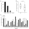
Figure 1
Figure 1
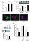
Wnt11 expression in human HCC tissue samples and cell lines. A, The level of Wnt11 mRNA was measured in 4 different HCC cell lines (HepG2, Hep3B, Huh7, and FOCUS) and plotted as copy numbers. No detectable expression of Wnt11 mRNA was found in FOCUS cells. B, Seventeen paired human HCC and corresponding adjacent HCC-free tissue were analyzed for Wnt11 mRNA expression. Wnt11 was significantly downregulated in HCC tumors compared to the adjacent peritumoral liver tissue. Horizontal bars indicate the mean values within each group of samples. Statistical comparisons were made using paired t-tests (p=0.0177). C, The level of Wnt11 mRNA plotted as a bar graph. The black bars represent the mRNA levels in HCC tissues, and the white bars in corresponding peritumoral areas. Within the paired samples, 11 of 17 (65%) showed decreased expression of Wnt11 mRNA in tumors compared with corresponding peritumoral tissues. D, Western blot analysis of Wnt11 expression in human HCC. Expression of Wnt11 protein was detected by anti-Wnt11 antibody and actin was used as a loading control (bottom panel). The level of Wnt11 protein was plotted as a ratio to actin (top panel). Note that 8 of 10 samples exhibited reduced expression level in tumor (T) compared to peritumoral (pT) tissues. E, Wnt11 protein expression in human HCC tissue samples using immunohistochemical statining. Representative example (case #5) of HCC and pertitumoral area was immunostained with anti-Wnt11 antibody (brown color) and counterstained with hematoxylin (blue color). Weak but clear positive signal for Wnt11 was found in the cytoplasm of hepatocytes in the peritumor liver tissue (right). In contrast, the immunoreactive Wnt11 in the HCC tissue was negative (left). (Magnification, × 100)
Figure 1
Figure 1
Wnt11 expression in human HCC tissue samples and cell lines. A, The level of Wnt11 mRNA was measured in 4 different HCC cell lines (HepG2, Hep3B, Huh7, and FOCUS) and plotted as copy numbers. No detectable expression of Wnt11 mRNA was found in FOCUS cells. B, Seventeen paired human HCC and corresponding adjacent HCC-free tissue were analyzed for Wnt11 mRNA expression. Wnt11 was significantly downregulated in HCC tumors compared to the adjacent peritumoral liver tissue. Horizontal bars indicate the mean values within each group of samples. Statistical comparisons were made using paired t-tests (p=0.0177). C, The level of Wnt11 mRNA plotted as a bar graph. The black bars represent the mRNA levels in HCC tissues, and the white bars in corresponding peritumoral areas. Within the paired samples, 11 of 17 (65%) showed decreased expression of Wnt11 mRNA in tumors compared with corresponding peritumoral tissues. D, Western blot analysis of Wnt11 expression in human HCC. Expression of Wnt11 protein was detected by anti-Wnt11 antibody and actin was used as a loading control (bottom panel). The level of Wnt11 protein was plotted as a ratio to actin (top panel). Note that 8 of 10 samples exhibited reduced expression level in tumor (T) compared to peritumoral (pT) tissues. E, Wnt11 protein expression in human HCC tissue samples using immunohistochemical statining. Representative example (case #5) of HCC and pertitumoral area was immunostained with anti-Wnt11 antibody (brown color) and counterstained with hematoxylin (blue color). Weak but clear positive signal for Wnt11 was found in the cytoplasm of hepatocytes in the peritumor liver tissue (right). In contrast, the immunoreactive Wnt11 in the HCC tissue was negative (left). (Magnification, × 100)
Figure 2
Figure 2
Over-expression of Wnt11 inhibits canonical Wnt signaling. A, Effects of Wnt11 on TCF transcriptional activity. The TCF activity was significantly decreased in FOCUS and Huh7 cells by 68% and 50%, respectively, but not in HepG2 cells which has an inactivating β-catenin mutation. B, Effect of Wnt11 on β-catenin nuclear accumulation in Huh7 cells by a double-immunofluoescence staining. Nuclear accumulation of β-catenin was reduced in Wnt11-expressed cells (bottom, white arrows), whereas control cells revealed high level of β-catenin accumulation in the nucleus (top, yellow arrows). C, Over-expression of Wnt11 decreased β-catenin levels in the manner of GSK-3β-independent. Over-expression of Wnt11 reduced total β-catenin levels with a concomitant increase in phospho-β-catenin; however, there was no difference between total GSK-3β and phospho-GSK-3β level. The bar graphs depict the results of densitometric analysis from the Western blots (bottom). The results are reported as the mean of three independent experiments. *: p < 0.05 D, Effect of GSK-3β inhibitor (LiCl) on β-catenin-dependent TCF activity in Wnt11 overexpressing cells. LiCl treatment did not rescue the reduced TCF activity observed in Wnt11 overexpressing FOCUS cells (W11-myc). E, Reduced cell proliferation in Wnt11 over-expressing cells. Cell proliferation rate was significantly decreased in FOCUS (FW11-1 and FW11-2) compared to control cells (F-C). Constitutive Wnt11 over-expression was demonstrated by Western blot analysis (top panel). *: p < 0.05 versus control cells
Figure 3
Figure 3
Wnt11 expression activated PKC signaling. A, Effect of Wnt11 on intracellular Ca2+, which was significantly increased in Wnt11 over-expressing (FW11-1, FW11-2) compare to control (F-C) cells. B, Effect of Wnt11 on PKC activity. Bar graph represents relative optical densities that reflect enzymatic activities. PKC activities are increased by 19% in FOCUS and 28% in Huh7 cells. C, Effect of Wnt11 on cellular distribution of PKC in FOCUS cells. Yellow arrows indicate translocation of activated PKC to the plasma membrane as visualized by FITC (green)- and Alexa594 (red)- conjugated secondary antibodies. D, Knockdown of Wnt11 expression with siRNA resulted in decreased PKC activity in Hep3B cells. (Top panel) Immunoblot analysis to verify knockdown of endogenous Wnt11 protein expression. Control siRNA (C-si) or Wnt11-siRNA (W11-si) was transfected into Hep3B cells. Ectopic expression of Wnt11-myc (W11-myc) was used as a positive control. Samples were immunoblotted with anti-Wnt11 antibody to detect endogenous Wnt11 protein. (Bottom panel) PKC activity was decreased in Wnt11-siRNA transfected Hep3B cells. E, The effect of PKC (BisI) and ROCK (Y27632) inhibitors on TCF activity measured after 24 h incubation with BisI or Y27632. BisI rescued the inhibition of TCF activity as mediated by Wnt11. In contrast, Y27632 had no effect.
Figure 4
Figure 4
Effects of Wnt11 on cell migration. A, Morphological alterations in a stable transfected Wnt11 over-expressing FOCUS (FW11-1). FW11-1 cells reveal a smooth leading edge with few membrane protrusions compared to control cells (left panel). Control and FW11-1 were subjected to immunostaining for F-actin with rhodamine-phalloidin (red, right panel). Note that control cells are characterized by thin stress fiber at the leading front edge of cells, whereas Wnt11 over-expressing cells show many bundled stress fiber without preferred orientation and suggest a less motile phenotype. B, Delayed cell migration in stable Wnt11 over-expressing clones compared to control cells using a wound-healing assay. The cells were photographed at the identical location at the time indicated. At 40 h, most of the wound was closed with migrating cells in the controls (F-C), whereas it remained open in Wnt11 over-expressing cells (FW11-1). Graph of the wound closure plotted against time. * p < 0.05, ** p < 0.001 versus control. C, Wnt11 inhibited cell motility using a Transwell chamber assay. (Top panel) Cell migration in Wnt11 over-expressing cells (FW11-1, FW11-2) was significantly reduced by 40 % compared to control (F-C). (Bottom panel) A representative cell motility assay stained with crystal violet. Number of migrated cells was decreased in FW11-1 compared to control. D, The Wnt11-mediated inhibition of cell motility was partially rescued by Wnt11 siRNA (W11-si). Knockdown of Wnt11 expression with siRNA was confirmed by Western blot analysis (top panel). E. Knockdown of Wnt11 resulted in increased cell motility in Hep3B cells.
Figure 5
Figure 5
Wnt11 decreased FOCUS cell motility through the RhoA/ROCK and Rac1 pathway. A, Wnt11 induced activation of RhoA/ROCK in FOCUS and Huh7 cells. Whole cell lysates were used to pull-down the active form of RhoA (GTP-RhoA) with GST-human rhotekin and followed by Western blot analysis. Equal amount of total cell lysates serve as controls (total RhoA). The bar graph shows the ratio of active RhoA to total RhoA, and ROCK2 to actin. B, Wnt11 decreased Rac1 activity in FOCUS cells. Wnt11 over-expresssing cells, either transiently (W11-myc) or stably (FW11-1, FW11-2) exhibited reduced Rac1 activity assessed by pull-down assay with GST-human Pak1 to detect active Rac1 (GTP-Rac1) using Western blot analysis. Aliquots of the respective lysates serve as controls for analyzing total amount of Rac1 protein (total Rac1). C, ROCK inhibitor (Y27632) rescued the decreased Rac1 activity mediated by Wnt11. Wnt11 over-expressing or control cells were treated with Y27632, followed by Rac1 activity assay. Y27632 restored the inhibition of Rac1 activity medicated by Wnt11, indicating that ROCK is responsible for the reduced Rac1 activity. D, Inhibition of cell motility was restored by ROCK inhibitors (HA1077, Y27632), not by BisI.
Figure 6
Figure 6
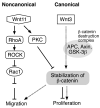
Schematic diagram of the potential crosstalk between canonical and noncanonical pathways as mediated by Wnt11 in HCC cells. Activation of the canonical Wnt signaling induces cell proliferation and migration through β-catenin stabilization and translocation to the nucleus where it activates Wnt responsive target genes (dotted arrows). The activation of PKC by Wnt11 triggers β-catenin phosphorylation and results in inhibition of cell proliferation. Wnt11-mediated activation of RhoA/ROCK inhibits Rac1 activity, and leads to inhibition of cell migration and motility (black arrows). Both Wnt3 (4) and Wnt11 may be involved in tumorigenesis by reciprocal mechanisms. Thus, over-expression of Wnt3 or down-regulation of Wnt11 or both may activate canonical β-catenin signaling and contribute to a highly motile, invasive and proliferative HCC phenotype. Therefore, a balance between Wnt3/canonical and Wnt11/noncanonical signaling may be important for homeostatic regulation of β-catenin signaling in liver and alterations in this balance may contribute to hepatic oncogenesis.

Similar articles

Cited by

References

    1. Laurent-Puig P, Legoix P, Bluteau O, et al. Genetic alterations associated with hepatocellular carcinomas define distinct pathways of hepatocarcinogenesis. Gastroenterology. 2001;120:1763–73. - PubMed
    1. Merle P, de la Monte S, Kim M, et al. Functional consequences of Frizzled-7 receptor overexpression in human hepatocellular carcinoma. Gastroenterology. 2004;127:1110–22. - PubMed
    1. Merle P, Kim M, Herrmann M, et al. Oncogenic role of the frizzled-7/β-catenin pathway in hepatocellular carcinoma. J Hepatol. 2005;43:854–62. - PubMed
    1. Kim M, Lee H, Tsedensodnom O, et al. Functional interaction between Wnt3 and Frizzled-7 leads to activation of the Wnt/beta-catenin signaling pathway in hepatocelluar carcinoma cells. J Hepatol. 2008;48:780–91. - PMC - PubMed
    1. Logan CY, Nusse R. The Wnt signaling pathway in development and disease. Annu Rev Cell Dev Biol. 2004;20:781–810. - PubMed

Publication types

MeSH terms