

Heterogeneity for stem cell-related markers according to tumor subtype and histologic stage in breast cancer
- PMID: 20103682
- PMCID: PMC2818503
- DOI: 10.1158/1078-0432.CCR-09-1532
Heterogeneity for stem cell-related markers according to tumor subtype and histologic stage in breast cancer
Abstract
Purpose: To evaluate the expression of stem cell-related markers at the cellular level in human breast tumors of different subtypes and histologic stage.
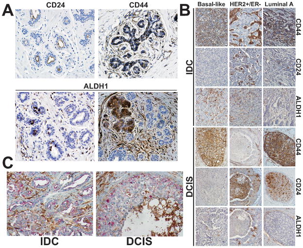
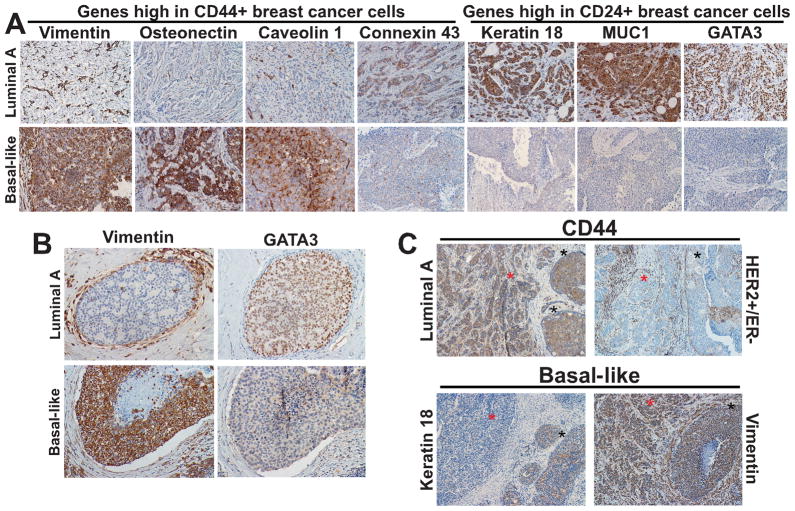
Experimental design: We performed immunohistochemical analyses of 12 proteins [CD44, CD24, ALDH1, vimentin, osteonectin, EPCR, caveolin 1, connexin 43, cytokeratin 18 (CK18), MUC1, claudin 7, and GATA3] selected based on their differential expression in breast cancer cells with more differentiated and stem cell-like characteristics in 47 cases of invasive ductal carcinoma (IDC) only, 135 cases of IDC with ductal carcinoma in situ (DCIS), 35 cases of DCIS with microinvasion, and 58 cases of pure DCIS. We also analyzed 73 IDCs with adjacent DCIS to determine the differences in the expression of markers by histology within individual tumors. CD44+/CD24- and CD24-/CD24+ cells were detected using double immunohistochemistry.
Results: CD44 and EPCR expression was different among the four histologic groups and was lower in invasive compared with in situ tumors, especially in luminal A subtype. The expression of vimentin, osteonectin, connexin 43, ALDH1, CK18, GATA3, and MUC1 differed by tumor subtype in some histologic groups. ALDH1-positive cells were more frequent in basal-like and HER2+ than in luminal tumors. CD44+/CD24- cells were detected in 69% of all tumors with 100% of the basal-like and 52% of HER2+ tumors having some of these cells.
Conclusions: Our findings suggest that in breast cancer, the frequency of tumor cells positive for stem cell-like and more differentiated cell markers varies according to tumor subtype and histologic stage.
Figures
Similar articles
-
ALDH1 is a better clinical indicator for relapse of invasive ductal breast cancer than the CD44+/CD24- phenotype.Med Oncol. 2014 Mar;31(3):864. doi: 10.1007/s12032-014-0864-0. Epub 2014 Feb 12. Med Oncol. 2014. PMID: 24519209
-
Unraveling the roles of CD44/CD24 and ALDH1 as cancer stem cell markers in tumorigenesis and metastasis.Sci Rep. 2017 Oct 23;7(1):13856. doi: 10.1038/s41598-017-14364-2. Sci Rep. 2017. PMID: 29062075 Free PMC article.
-
Clinical-pathologic significance of cancer stem cell marker expression in familial breast cancers.Breast Cancer Res Treat. 2013 Jul;140(1):195-205. doi: 10.1007/s10549-013-2591-1. Epub 2013 Jun 28. Breast Cancer Res Treat. 2013. PMID: 23813303 Free PMC article.
-
Breast cancer stem cells and intrinsic subtypes: controversies rage on.Curr Stem Cell Res Ther. 2009 Jan;4(1):50-60. doi: 10.2174/157488809787169110. Curr Stem Cell Res Ther. 2009. PMID: 19149630 Review.
-
Mammary stem cells and breast cancer--role of Notch signalling.Stem Cell Rev. 2007 Jun;3(2):169-75. doi: 10.1007/s12015-007-0023-5. Stem Cell Rev. 2007. PMID: 17873349 Review.
Cited by
-
Efficacy of using cancer stem cell markers in isolating and characterizing liver cancer stem cells.Stem Cells Dev. 2013 Oct 1;22(19):2655-64. doi: 10.1089/scd.2012.0703. Epub 2013 Jun 8. Stem Cells Dev. 2013. PMID: 23638793 Free PMC article.
-
Breast cancer stem-like cells: clinical implications and therapeutic strategies.Clujul Med. 2016;89(2):193-8. doi: 10.15386/cjmed-559. Epub 2016 Apr 15. Clujul Med. 2016. PMID: 27152067 Free PMC article. Review.
-
Kruppel-like factor 4 signals through microRNA-206 to promote tumor initiation and cell survival.Oncogenesis. 2015 Jun 8;4(6):e155. doi: 10.1038/oncsis.2015.8. Oncogenesis. 2015. PMID: 26053033 Free PMC article.
-
The bed and the bugs: interactions between the tumor microenvironment and cancer stem cells.Semin Cancer Biol. 2012 Oct;22(5-6):462-70. doi: 10.1016/j.semcancer.2012.04.006. Epub 2012 Apr 23. Semin Cancer Biol. 2012. PMID: 22548722 Free PMC article. Review.
-
Relationship of CD44+CD24-/low breast cancer stem cells and axillary lymph node metastasis.J Transl Med. 2012 Sep 19;10 Suppl 1(Suppl 1):S6. doi: 10.1186/1479-5876-10-S1-S6. Epub 2012 Sep 19. J Transl Med. 2012. PMID: 23046710 Free PMC article.
References
Publication types
MeSH terms
Substances
Grants and funding
LinkOut - more resources
Full Text Sources
Other Literature Sources
Medical
Research Materials
Miscellaneous
