

HOXA10 regulates endometrial GABAA {pi} receptor expression and membrane translocation
- PMID: 20103740
- PMCID: PMC3774337
- DOI: 10.1152/ajpendo.00577.2009
HOXA10 regulates endometrial GABAA {pi} receptor expression and membrane translocation
Abstract
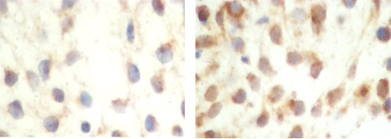
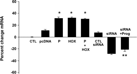
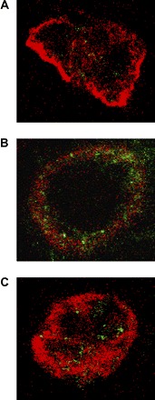
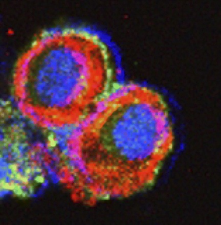
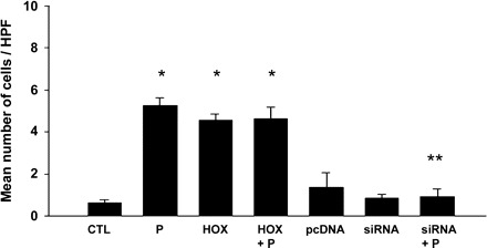
Expression of the GABA(A) pi receptor has been described previously in the human endometrium in both luminal epithelium and stroma. Its expression is increased during decidualization in rodents and in the implantation window of human endometrium. Here we localized GABA pi subunit receptor protein in human endometrium and identified regulators of gene expression and activation. GABA(A) pi was localized to the cell surface, and expression increased during the window of embryo implantation in human endometrium. The well-differentiated human endometrial adenocarcinoma cell line Ishikawa was treated with progesterone and transfected with pcDNA-HOXA10, HOXA10 siRNA, or respective controls. GABA(A) pi receptor mRNA expression was evaluated by real-time RT-PCR. Protein expression and localization were evaluated using immunofluorescence. GABA(A) pi receptor mRNA expression was increased significantly after either progesterone treatment or HOXA10 transfection. Coadministration of progesterone along with HOXA10 transfection had no additional effect on the expression of GABA(A) pi receptor mRNA over either agent alone. Blocking HOXA10 expression with siRNA prevented progesterone-induced GABA(A) pi receptor mRNA expression. Additionally, either HOXA10 or progesterone independently caused increased translocation of the GABA receptor from the cytoplasm to the cell membrane. Translocation in response to progesterone was blocked with HOXA10 siRNA. Progesterone-induced GABA(A) pi subunit receptor expression is likely mediated indirectly through progesterone's regulation of HOXA10 expression. Modification of subtype composition and translocation of the GABA receptor ion channel likely modulate endometrial receptivity. Whereas HOXA10 typically enhances the expression of progesterone-responsive genes, here HOXA10 expression leads to production of a less progestin-responsive GABA receptor subtype, likely buffering the effects of luteal phase progesterone on GABA receptor activity.
Figures
Similar articles
-
Endothelin type A receptor (ETA) expression is regulated by HOXA10 in human endometrial stromal cells.Reprod Sci. 2010 May;17(5):471-6. doi: 10.1177/1933719110361961. Reprod Sci. 2010. PMID: 20371740 Free PMC article.
-
HOXA10 inhibits Kruppel-like factor 9 expression in the human endometrial epithelium.Biol Reprod. 2010 Aug 1;83(2):205-11. doi: 10.1095/biolreprod.110.083980. Epub 2010 May 12. Biol Reprod. 2010. PMID: 20463357 Free PMC article.
-
Regulation of homeobox A10 expression in the primate endometrium by progesterone and embryonic stimuli.Reproduction. 2007 Sep;134(3):513-23. doi: 10.1530/REP-07-0234. Reproduction. 2007. PMID: 17709569
-
Role of HOXA10 in pathologies of the endometrium.Rev Endocr Metab Disord. 2025 Feb;26(1):81-96. doi: 10.1007/s11154-024-09923-8. Epub 2024 Nov 5. Rev Endocr Metab Disord. 2025. PMID: 39499452 Review.
-
Homeobox genes in endometrium: from development to decidualization.Int J Dev Biol. 2020;64(1-2-3):227-237. doi: 10.1387/ijdb.190120dm. Int J Dev Biol. 2020. PMID: 32659011 Review.
Cited by
-
Bioinformatic analysis of endometrial miRNA expression profile at day 26-28 of pregnancy in the mare.Sci Rep. 2024 Feb 16;14(1):3900. doi: 10.1038/s41598-024-53499-x. Sci Rep. 2024. PMID: 38365979 Free PMC article.
-
GABA(A) receptor pi (GABRP) stimulates basal-like breast cancer cell migration through activation of extracellular-regulated kinase 1/2 (ERK1/2).J Biol Chem. 2014 Aug 29;289(35):24102-13. doi: 10.1074/jbc.M114.593582. Epub 2014 Jul 10. J Biol Chem. 2014. PMID: 25012653 Free PMC article.
-
Characterization of the Transcriptional Complexity of the Receptive and Pre-receptive Endometria of Dairy Goats.Sci Rep. 2015 Sep 16;5:14244. doi: 10.1038/srep14244. Sci Rep. 2015. PMID: 26373443 Free PMC article.
-
GABA A receptor π subunit promotes apoptosis of HTR-8/SVneo trophoblastic cells: Implications in preeclampsia.Int J Mol Med. 2016 Jul;38(1):105-12. doi: 10.3892/ijmm.2016.2608. Epub 2016 May 25. Int J Mol Med. 2016. PMID: 27221053 Free PMC article.
-
GABRP is a potential prognostic biomarker and correlated with immune infiltration and tumor microenvironment in pancreatic cancer.Transl Cancer Res. 2022 Apr;11(4):649-668. doi: 10.21037/tcr-21-2021. Transl Cancer Res. 2022. PMID: 35571651 Free PMC article.
References
-
- Andriamampandry C, Taleb O, Kemmel V, Humbert JP, Aunis D, Maitre M. Cloning and functional characterization of a gamma-hydroxybutyrate receptor identified in the human brain. FASEB J 21: 885–895, 2007 - PubMed
-
- Apud JA, Tappaz ML, Celotti F, Negri-Cesi P, Masotto C, Racagni G. Biochemical and immunochemical studies on the GABAergic system in the rat fallopian tube and ovary. J Neurochem 43: 120–125, 1986 - PubMed
-
- Bagot CN, Troy PJ, Taylor HS. Alteration of maternal Hoxa10 expression by in vivo gene transfection affects implantation. Gene Ther 7: 1378–1384, 2000 - PubMed
-
- Benson GV, Lim H, Paria BC, Satokata I, Dey SK, Maas RL. Mechanisms of reduced fertility in Hoxa-10 mutant mice: uterine homeosis and loss of maternal Hoxa-10 expression. Development 122: 2687–2696, 1996 - PubMed
-
- Castelbaum AJ, Ying L, Somkuti SG, Sun J, Ilesanmi AO, Lessey BA. Characterization of integrin expression in a well differentiated endometrial adenocarcinoma cell line (Ishikawa). J Clin Endocrinol Metab 82: 136–142, 1997 - PubMed
Publication types
MeSH terms
Substances
Grants and funding
LinkOut - more resources
Full Text Sources
Research Materials
Miscellaneous