Review
. 2010 Mar 2;102(5):789-95.
doi: 10.1038/sj.bjc.6605551. Epub 2010 Jan 26.

Hypoxia inducible factors in cancer stem cells

Affiliations
Review

Hypoxia inducible factors in cancer stem cells

J M Heddleston et al. Br J Cancer. .

Abstract

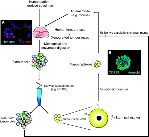
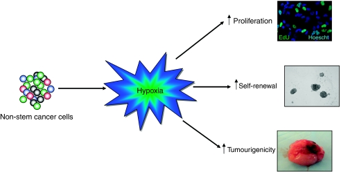
Oxygen is an essential regulator of cellular metabolism, survival, and proliferation. Cellular responses to oxygen levels are monitored, in part, by the transcriptional activity of the hypoxia inducible factors (HIFs). Under hypoxia, HIFs regulate a variety of pro-angiogenic and pro-glycolysis pathways. In solid cancers, regions of hypoxia are commonly present throughout the tissue because of the chaotic vascular architecture and regions of necrosis. In these regions, the hypoxic state fluctuates in a spatial and temporal manner. Transient hypoxic cycling causes an increase in the activity of the HIF proteins above what is typical for non-pathologic tissue. The extent of hypoxia strongly correlates to poor patient survival, therapeutic resistance and an aggressive tumour phenotype, but the full contribution of hypoxia and the HIFs to tumour biology is an area of active investigation. Recent reports link resistance to conventional therapies and the metastatic potential to a stem-like tumour population, termed cancer stem cells (CSCs). We and others have shown that within brain tumours CSCs reside in two niches, a perivascular location and the surrounding necrotic tissue. Restricted oxygen conditions increase the CSC fraction and promote acquisition of a stem-like state. Cancer stem cells are critically dependant on the HIFs for survival, self-renewal, and tumour growth. These observations and those from normal stem cell biology provide a new mechanistic explanation for the contribution of hypoxia to malignancy. Further, the presence of hypoxia in tumours may present challenges for therapy because of the promotion of CSC phenotypes even upon successful killing of CSCs. The current experimental evidence suggests that CSCs are plastic cell states governed by microenvironmental conditions, such as hypoxia, that may be critical for the development of new therapies targeted to disrupt the microenvironment.


Figures

Figure 1
Figure 1
Enrichment of cultures for cancer stem cells allow for better study of their unique biology. In order to appropriately examine the biological significance of the cancer stem cell population, in vitro cultures must be enriched for this population before experimental investigation. Utilising animal models, such as immunocompromised mice, patient-derived cancer cells can be expanded for use in the laboratory. Following resection of the tumour from the patient, the mass is dissociated into single cells through a combination of mechanical and enzymatic digestion. Once the cells have recovered and are growing as single cells, they can be sorted based on surface marker expression. Experimental evidence has demonstrated that the cancer stem cell sub-population express a subset of genes that can act as markers for enriching cultures for the stem-like cancer cells (Singh et al, 2003). The present gold standard of these markers in glioma is the surface protein, CD133 (also known as Prominin-1). CD133 can be used in cell sorting to discern the cancer stem cell sub-population. Although little is known about its role in cancer biology, (A) CD133 expression has been observed on a sub-population of cells in the tumour tissue and (B) this expression is maintained in the cancer stem cell population in vitro. In (A) a subcutaneous tumour was resected out of a mouse, fixed in paraformaldehyde, and frozen. Sections of the tumour were cut and stained for CD133 (red) and nuclei (Hoescht 3342, blue). In (B) cancer stem cells enriched from glioblastoma multiforme were cultured in serum-free media until they formed spheroids (neurospheres). The cells were then fixed, frozen, and cut. The sections were stained for CD133 (green) and nuclei (Hoescht 3342, blue). This procedure allowed for direct staining of cells throughout the sphere. Once the tumour cells have been separated into cancer stem cell (CSC)-enriched and non-CSC populations, experiments can be carried out to elucidate the biological differences between the two populations. The most important way to test CSC biology is the ability of the cancer stem cells to form tumours that are phenotypic copies of the parental tumour. In the glioma system, one way to determine a cells tumourigenic capacity is direct intracranial implantation. In this way, the tumour formation capacity of cancer cells allows researchers to delineate the stem-like fraction of cells within a tumour.
Figure 2
Figure 2
Hypoxia and the hypoxia inducible factors (HIFs) can promote the stem-like phenotype. Recent experimental evidence has demonstrated that HIFs have crucial roles in cancer. Following culture in low-oxygen conditions, the phenotype non-stem cancer cells were pushed to a more stem-like state. Several important characteristics of the cancer stem cell population, such as enhanced growth, self renewal (evidenced by spheroid formation), and tumourigenesis, have been shown to increase in the non-stem fraction of cancer cells following hypoxic culture (Heddleston et al, 2009). Non-stem cells increase their rate of proliferation above what is normally seen at 20% oxygen. This can be visualised by EdU retention (an analogue of BrDU). Hypoxia has also been shown to promote self-renewal, which can be measured by spheroid growth starting from a single cell. Non-stem cancer cells expressing constitutively active HIF2α protein have been demonstrated to have increased tumourigenicity in mice animal models.
Figure 3
Figure 3
A new paradigm in modelling tumour biology. Classically cancer cell biology has been modelled in a stochastic or hierarchical sense. These models focus on which cells within a tumour population are responsible for tumourigenicity, but treat cellular progression an irreversible process: either all (or a large majority) can form tumours even as the cell phenotype may change, or that only the parental cells of the hierarchy (cancer stem cells) can form tumours, whereas their differentiated progeny act as a support structure. A new way to view the cellular organisation of tumour is to focus on how a cell may acquire the ability to be tumourigenic, rather than what definitive cell population forms the tumour. The mutation model posits that the ability to form tumours is because of an accumulation of specific mutations that allow the cells to evade therapy and growth more rapidly. In this model, each mutation is irreversible and the accumulation of important mutations (e.g. p53, EGFR) allows for a sustained sub-population of cancer stem cells. The second model describes the cellular structure of the tumour to be more plastic and allow for cellular adaptation to the microenvironment. The cancer stem cell sub-population drives tumour growth, but this population arises from adaptation of the cells to their microenvironment. External influences, such as hypoxia, can drive a reversible phenotype that can enhance stem-like properties of cells to ensure survival of the tumour.

Similar articles

Cited by

References

    1. Al-Hajj M, Wicha MS, Benito-Hernandez A, Morrison SJ, Clarke MF (2003) Prospective identification of tumorigenic breast cancer cells. Proc Natl Acad Sci USA 100: 3983–3988 - PMC - PubMed
    1. Baish JW, Jain RK (1998) Cancer, angiogenesis and fractals. Nat Med 4: 984 - PubMed
    1. Bao S, Wu Q, McLendon RE, Hao Y, Shi Q, Hjelmeland AB, Dewhirst MW, Bigner DD, Rich JN (2006a) Glioma stem cells promote radioresistance by preferential activation of the DNA damage response. Nature 444: 756–760 - PubMed
    1. Bao S, Wu Q, Sathornsumetee S, Hao Y, Li Z, Hjelmeland AB, Shi Q, McLendon RE, Bigner DD, Rich JN (2006b) Stem cell-like glioma cells promote tumor angiogenesis through vascular endothelial growth factor. Cancer Res 66: 7843–7848 - PubMed
    1. Ben-Porath I, Thomson MW, Carey VJ, Ge R, Bell GW, Regev A, Weinberg RA (2008) An embryonic stem cell-like gene expression signature in poorly differentiated aggressive human tumors. Nat Genet 40: 499–507 - PMC - PubMed

Publication types

Substances