. 2010 Feb;31(3):478-90.
doi: 10.1111/j.1460-9568.2010.07077.x. Epub 2010 Jan 25.

Impact of dendritic spine preservation in medium spiny neurons on dopamine graft efficacy and the expression of dyskinesias in parkinsonian rats

Affiliations

Impact of dendritic spine preservation in medium spiny neurons on dopamine graft efficacy and the expression of dyskinesias in parkinsonian rats

Katherine E Soderstrom et al. Eur J Neurosci. 2010 Feb.

Abstract

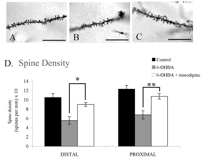
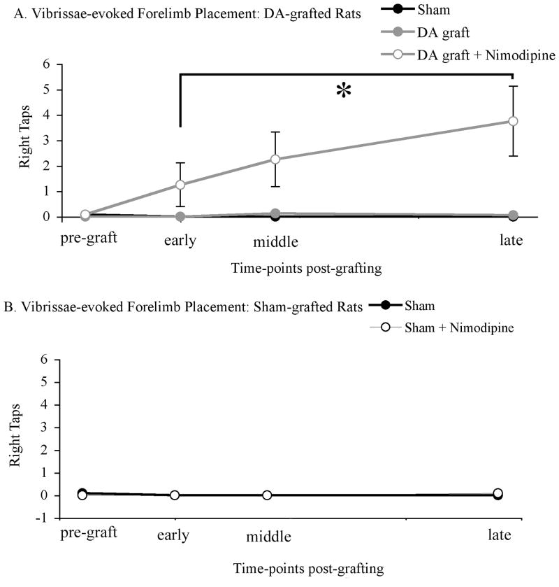
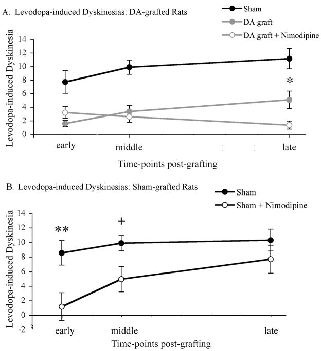
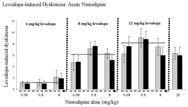
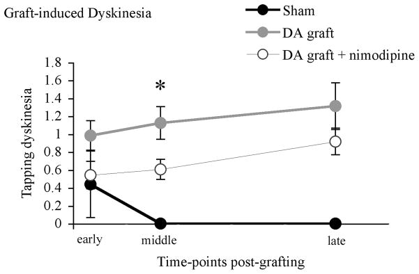
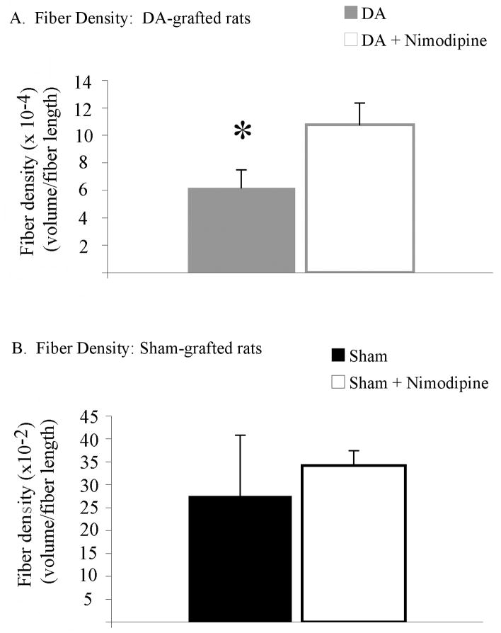
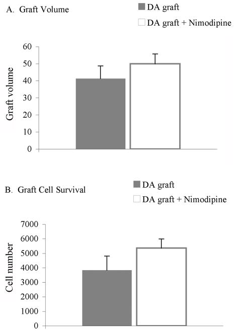
Dopamine deficiency associated with Parkinson's disease (PD) results in numerous changes in striatal transmitter function and neuron morphology. Specifically, there is marked atrophy of dendrites and dendritic spines on striatal medium spiny neurons (MSN), primary targets of inputs from nigral dopamine and cortical glutamate neurons, in advanced PD and rodent models of severe dopamine depletion. Dendritic spine loss occurs via dysregulation of intraspine Cav1.3 L-type Ca(2+)channels and can be prevented, in animal models, by administration of the calcium channel antagonist, nimodipine. The impact of MSN dendritic spine loss in the parkinsonian striatum on dopamine neuron graft therapy remains unexamined. Using unilaterally parkinsonian Sprague-Dawley rats, we tested the hypothesis that MSN dendritic spine preservation through administration of nimodipine would result in improved therapeutic benefit and diminished graft-induced behavioral abnormalities in rats grafted with embryonic ventral midbrain cells. Analysis of rotational asymmetry and spontaneous forelimb use in the cylinder task found no significant effect of dendritic spine preservation in grafted rats. However, analyses of vibrissae-induced forelimb use, levodopa-induced dyskinesias and graft-induced dyskinesias showed significant improvement in rats with dopamine grafts associated with preserved striatal dendritic spine density. Nimodipine treatment in this model did not impact dopamine graft survival but allowed for increased graft reinnervation of striatum. Taken together, these results demonstrate that even with grafting suboptimal numbers of cells, maintaining normal spine density on target MSNs results in overall superior behavioral efficacy of dopamine grafts.


Figures

Figure 1
Figure 1. Experimental groups and timeline
Rats were placed into treatment groups with differential MSN spine density: a sham-grafted group receiving cell-free media plus vehicle pellets (n=6), a sham-grafted group receiving cell-free media plus nimodipine pellets (n=6), a dopamine-grafted group receiving embryonic ventral mesencephalic cell grafts plus vehicle pellets (n=8), and a dopamine-grafted group receiving embryonic ventral mesencephalic cell grafts plus nimodipine pellets (n=6). All rats received unilateral nigrostriatal 6-OHDA lesions. All pellets were delivered 24 hours following lesion and were replaced every 20 days till the conclusion of the study. Rats received dopamine or sham grafts 4 weeks following lesion. Behavioral analyses were conducted 4 weeks following grafting and continued till the completion of the study. DA=dopamine.
Figure 2
Figure 2. Slow-release nimodipine pellets preserve spine density in parkinsonian rats
(AC) Photomicrographs of MSN dendrites spine density in (A) untreated controls, (B) dopamine-depleted (C) dopamine-depleted plus nimodipine treated groups. (D) Rats receiving intranigral 6-OHDA lesions showed a significant reduction in striatal spine density when compared to control rats at both distal (p=0.002) and proximal (p=0.001) dendritic sites. This loss of spine density was diminished significantly in rats receiving slow-release nimodipine pellets at both distal (*p=0.012) and proximal (**p=0.007) sites. Scale bars represent 60X.
Figure 3
Figure 3. Rotational Asymmetry
(A) Dopamine grafting, regardless of spine density, showed a near complete amelioration of rotational asymmetry. Both grafted groups showed a significant and near complete amelioration of levodopa-induced dyskinesias when compared to sham- grafted rats (*DA graft vs. sham graft, p=0.001; DA graft + nimodipine vs. sham graft, p=0.001). (B) Nimodipine treatment failed to amelioration levodopa-induced rotations in sham-grafted rats. DA=dopamine.
Figure 4
Figure 4. Preserved striatal spine density significantly improved vibrissae-induced forelimb placement in dopamine-grafted rats (A), but not sham-grafted rats (B)
(A) Dopamine-grafted rats receiving continuous nimodipine treatment showed significantly more vibrissae-induced forelimb placement when compared to rats receiving grafts alone (*p=0.024) and sham-grafted rats (p=0.026). (B) Nimodipine treatment had no effect on vibrissae-induced forelimb placement in sham-grafted parkinsonian rats. DA=dopamine.
Figure 5
Figure 5. Preserving spine density significantly reduced the occurrence of levodopa-induced dyskinesias in dopamine-grafted rats (A) and sham-grafted rats (B)
(A) Both dopamine-grafted groups showed a significant amelioration of levodopa-induced dyskinesias (p=0.001). However, dopamine grafts placed into parkinsonian rats with intact spine density (DA graft + nimodipine) showed a significant further reduction in dyskinesias at later post-graft time-points compared with parkinsonian rats grafted with dopamine neurons and experiencing dendritic spine loss (DA graft + vehicle, *p=0.02). (B) Preserving spine density also resulted in an amelioration of levodopa-induced dyskinesias in parkinsonian rats receiving sham grafts at early (**p=0.005) and middle (+p=0.013) time-points post-grafting, but not by the conclusion of the experiment (p=0.176). DA=dopamine.
Figure 6
Figure 6. Acute nimodipine treatment did not impact levodopa-induced dyskinesias
A group of parkinsonian rats, distinct from rats used for the chronic nimodipine pellet studies, were employed. Dyskinesia severity was analyzed at 30 minutes after levodopa, but prior to nimodipine (light gray bars). Immediately following this behavioral evaluation, dyskinetic rats were injected with 1 of 4 doses of nimodipine (0.08, 0.8, 8.0, or 20 mg/kg) and dyskinetic behaviors rated again, 30 minutes after nimodipine (black bars). Dashed arrows on x-axis indicate escalating doses of nimodipine tested in combination with 1 of 3 doses of levodopa (6.0, 8.0, or 12.5 mg/kg). The horizontal dashed lines indicate the statistical dyskinesia average for each dose of the 3 doses of levodopa used.
Figure 7
Figure 7. Preserving spine density did not significantly improve performance on the cylinder test
No difference was seen in right forelimb use in the cylinder task between groups (p=0.204). DA=dopamine.
Figure 8
Figure 8. Spine density preservation resulted in a transient improvement in the expression of the graft-induced dyskinesia, tapping
Continuous nimodipine treatment resulted in a significant decrease in the severity score of tapping dyskinesia in dopamine-grafted rats (DA graft + nimodipine) when compared with rats receiving dopamine-grafts alone (DA graft, *p=0.04) at a middle time-point post-grafting; however this effect was lost at later post-graft time-points (p=0.191). DA=dopamine.
Figure 9
Figure 9. Spine density preservation did result in an increase in graft outgrowth
(A) Despite having no effect on graft survival, continuous nimodipine treatment did result in an increase in TH+ fiber density in dopamine-grafted rats compared with rats receiving grafts alone (* p=0.05). (B) Preserving spine density had no effect on fiber density in sham-grafted rats. DA=dopamine.
Figure 10
Figure 10. Nimodipine pellets did not effect the survival of dopamine grafts
Continuous nimodipine treatment did not result in a difference in graft volume (p=0.371) or graft survival (p=0.219) between grafted groups. DA=dopamine.

Comment in

References

    1. Breysse N, Carlsson T, Winkler C, Björklund A, Kirik D. The functional impact of the intrastriatal dopamine neuron grafts in parkinsonian rats is reduced with advancing disease. 2007;27:5849–56. - PMC - PubMed
    1. Brown P, Gerfen CR. Plasticity within striatal direct pathway neurons after neonatal dopamine depletion is mediated through a novel functional coupling of serotonin 5-HT2 receptors to the ERK 1/2 map kinase pathway. J Comp Neurol. 2006;498:415–30. - PMC - PubMed
    1. Brundin P, Karlsson J, Emgard M, Schierle GS, Hansson O, Petersen A, Castilho RF. Improving the survival of grafted dopaminergic neurons: a review over current approaches. Cell Transplant. 2000;9:179–95. - PubMed
    1. Caligiuri MP, Lohr JB. Worsening of postural tremor in patients with levodopa- induced dyskinesia: a quantitative analysis. Clin Neuropharmacol. 1993;16:244–50. - PubMed
    1. Collier TJ, Lipton J, Daley BF, Palfi S, Chu Y, Sortwell C, Bakay RA, Sladek JR, Jr, Kordower JH. Aging-related changes in the nigrostriatal dopamine system and the response to MPTP in nonhuman primates: diminished compensatory mechanisms as a prelude to parkinsonism. Neurobiol Dis. 2007;26:56–65. - PMC - PubMed

Publication types

MeSH terms