. 2010 Jan 27:10:13.
doi: 10.1186/1471-213X-10-13.

Activated Ras alters lens and corneal development through induction of distinct downstream targets

Affiliations

Activated Ras alters lens and corneal development through induction of distinct downstream targets

Daniel Burgess et al. BMC Dev Biol. .

Abstract

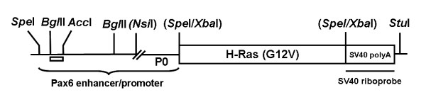
Background: Mammalian Ras genes regulate diverse cellular processes including proliferation and differentiation and are frequently mutated in human cancers. Tumor development in response to Ras activation varies between different tissues and the molecular basis for these variations are poorly understood. The murine lens and cornea have a common embryonic origin and arise from adjacent regions of the surface ectoderm. Activation of the fibroblast growth factor (FGF) signaling pathway induces the corneal epithelial cells to proliferate and the lens epithelial cells to exit the cell cycle. The molecular mechanisms that regulate the differential responses of these two related tissues have not been defined. We have generated transgenic mice that express a constitutively active version of human H-Ras in their lenses and corneas.

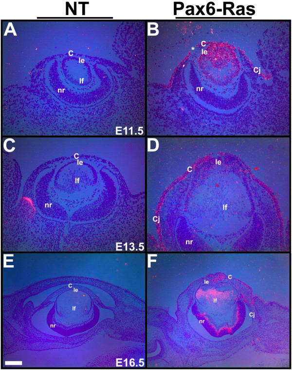
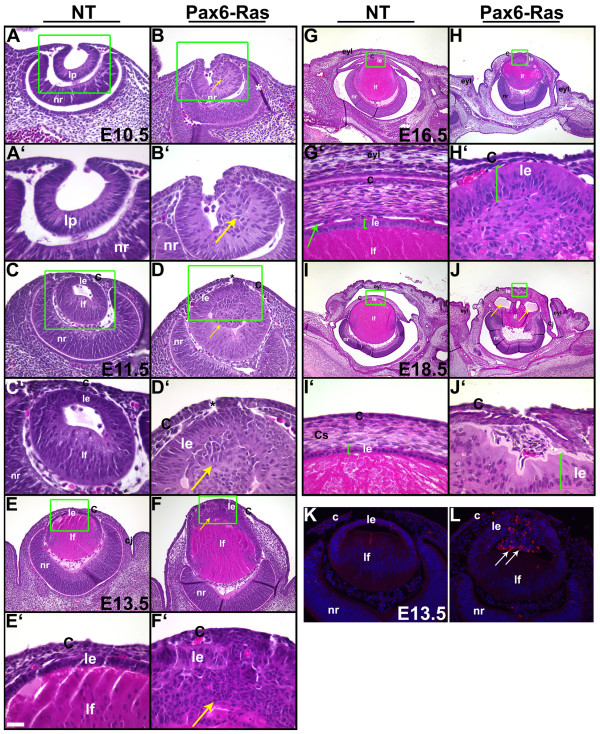
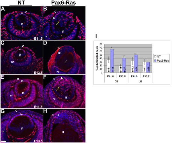
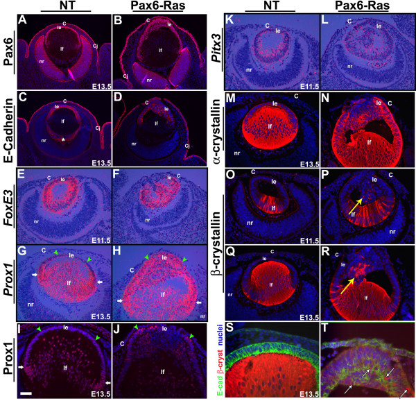
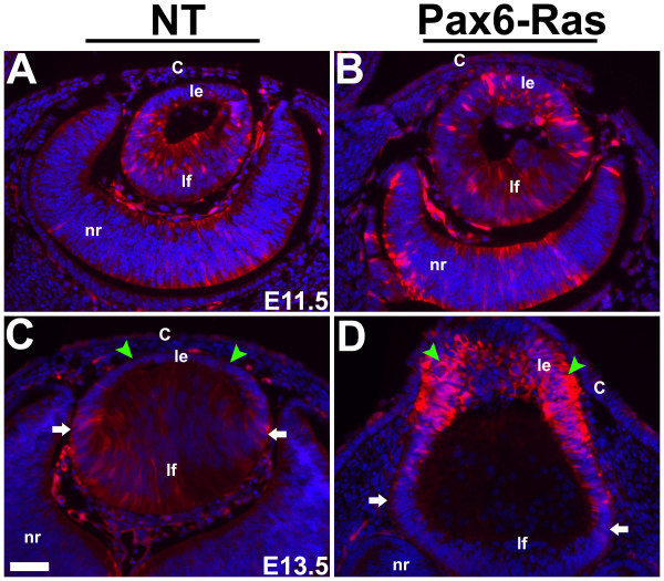
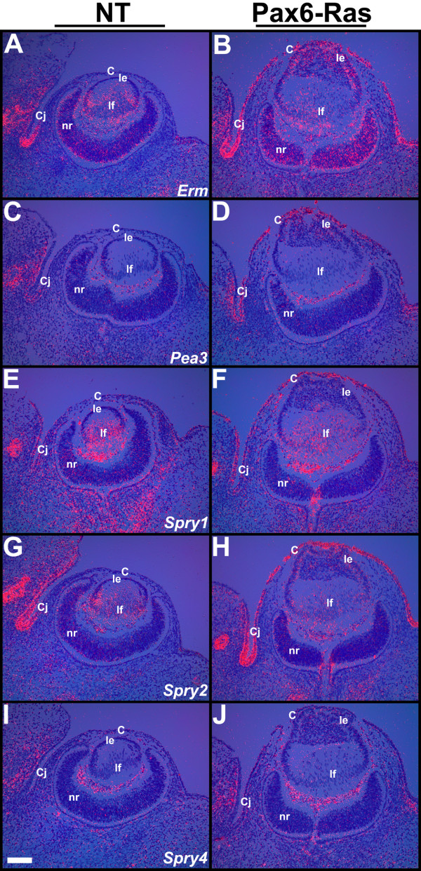
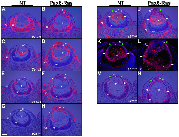
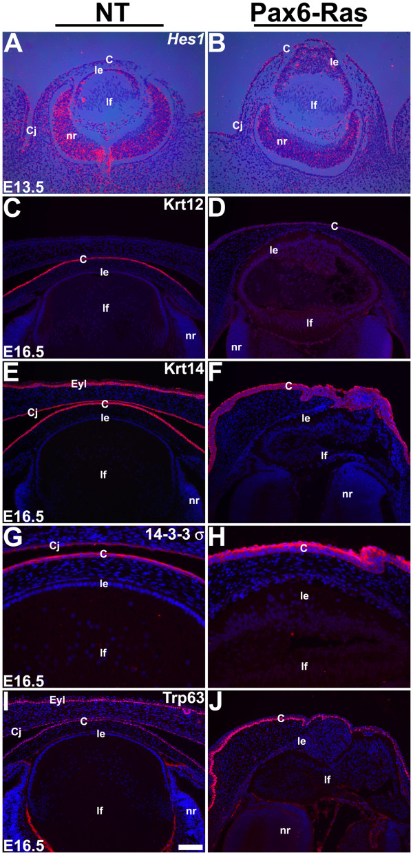
Results: Ras transgenic lenses and corneal epithelial cells showed increased proliferation with concomitant increases in cyclin D1 and D2 expression. This initial increase in proliferation is sustained in the cornea but not in the lens epithelial cells. Coincidentally, cdk inhibitors p27Kip1 and p57Kip2 were upregulated in the Ras transgenic lenses but not in the corneas. Phospho-Erk1 and Erk2 levels were elevated in the lens but not in the cornea and Spry 1 and Spry 2, negative regulators of Ras-Raf-Erk signaling, were upregulated more in the corneal than in the lens epithelial cells. Both lens and corneal differentiation programs were sensitive to Ras activation. Ras transgenic embryos showed a distinctive alteration in the architecture of the lens pit. Ras activation, though sufficient for upregulation of Prox1, a transcription factor critical for cell cycle exit and initiation of fiber differentiation, is not sufficient for induction of terminal fiber differentiation. Expression of Keratin 12, a marker of corneal epithelial differentiation, was reduced in the Ras transgenic corneas.

Conclusions: Collectively, these results suggest that Ras activation a) induces distinct sets of downstream targets in the lens and cornea resulting in distinct cellular responses and b) is sufficient for initiation but not completion of lens fiber differentiation.


Figures

Figure 1
Figure 1
Schematic diagram of the Pax6-Ras transgene. A human H-Ras (G12V) genomic clone (~3.1 Kb) was inserted between a modified Pax6 promoter/enhancer (2.3 Kb) and an intron and polyadenylation (polyA) sequence derived from the SV40 virus (850 bp). The lens/cornea enhancer is contained between the BglII/AccI sites (clear box). The SV40 polyA sequences were used to make riboprobes for detection of transgene expression.
Figure 2
Figure 2
Transgene expression. In situ hybridizations were performed on ocular sections of nontransgenic (A, C, E) and Pax6-Ras transgenic (B, D, F) embryos using an 35S-labeled SV40 riboprobe. Dark-field images were overlaid on respective bright-field images and silver grains were pseudocolored red. Ras transgene expression was seen in the lens, cornea and conjunctival epithelial cells at E11.5 and E13.5 (B, D) and in some retinal neuroblasts at E13.5 and E16.5 (D, F). Discontinuity in the corneal epithelium in panel B (asterisk) is due to a sectioning artifact. Abbreviations; C, corneal epithelium; Cj, conjunctival epithelium; le, lens epithelium; lf, lens fibers; nr, neural retina. The scale bar (E) represents 100 μm in panels A-D and 250 μm in panels E, F.
Figure 3
Figure 3
Pax6-Ras transgenic mice show altered lens and corneal development. Heads of E10.5 (A-B'), E11.5 (C-D'), E13.5 (E-F'), E16.5 (G-H') and E18.5 (I-J') nontransgenic (NT) and Pax6-Ras transgenic mice were sectioned and stained with hematoxylin and eosin. Panels A-J' are higher magnifications of boxed regions in A-J respectively. Asterisks in panels D, D' indicate sectioning artifacts. Arrows in B, B', D and D', point to abnormal clusters of cells in the interior of the lens. Arrow in G' points to the corneal endothelium. Arrows in J point to the vacuoles within the lens. Lens epithelial hyperplasia was seen in the Ras transgenic embryos (arrow in F') and Ras transgenic lenses failed to detach from the surface ectoderm (D, D'). Eyelids failed to close in Ras transgenic embryos (H) and the corneal stromal cells were disorganized (F, H, J). K, L. Activated caspase 3 was detected by immunohistochemistry (L, arrows). Antigen-antibody complexes are in red and the nuclei are stained blue with DAPI. Abbreviations; C, corneal epithelium; Cj, conjunctival epithelium; eyl, eyelid; le, lens epithelium; lf, lens fibers; lp, lens pit; nr, neural retina. The scale bar (E') represents 50 μm in panels A, B, C, D, K, L, 100 μm in panels E, F, 25 μm in A'-H' and 250 μm in panels G-J.
Figure 4
Figure 4
Cell proliferation in Ras transgenic mice. BrdU (A-D) and Ki67 (E-H) immunohistochemistry was performed on nontransgenic (NT) (A, C, E, G) and Pax6-Ras transgenic (B, D, F, H) embryos of age E11.5 (A, B, E, F) and E13.5 (C, D, G, H). Antigen antibody complexes are in red and nuclei are stained blue with DAPI. Quantification of the BrdU proliferation index is shown in panel I. Ras transgenic lens epithelial cells showed a significant increase in BrdU incorporation at E11.5 (I) but not at E13.5 (I). The number of sections counted for each genotype and time point are indicated in the graph. Error bars indicate standard deviation. Asterisk in the graph indicates a significance of P < 0.05. The scale bar (G) represents 50 μm in panels A, B, E-H and 100 μm in panels C, D.
Figure 5
Figure 5
Altered lens differentiation in Ras transgenic embryos. Immunohistochemistry (A-D, I, J, M-T) and in situ hybridizations (E-H, K, L) were performed on nontransgenic (NT) and Pax6-Ras transgenic embryos to detect expression of Pax6 (A, B), E-Cadherin (C, D, S, T), FoxE3 (E, F), Prox1 (G-J), Pitx3 (K, L), α-crystallin (M, N) and β-crystallin (O-T). In panels A-D, I, J and M-R, antigen antibody complexes are in red and in panels S and T, green and red. In all these panels nuclei are stained blue with DAPI. In situ hybridizations were performed using 35S-labeled riboprobes (E-H, K, L). Dark-field images were overlaid on respective bright-field images and silver grains were pseudocolored red. Upregulation of Prox1 (H, J) expression in a subset of Ras transgenic lens epithelial cells suggests initiation of early fiber differentiation. Co-localization of β-crystallin and E-Cadherin was seen in some of the Ras transgenic lens cells (T, arrows). The staining in the vitreous (panel C, asterisk) is due to anti-mouse secondary antibody binding to IgGs in the blood vessels. White arrows in panel G, H and I point to the lens equatorial region where fiber differentiation is initiated. Green arrowheads point to lens epithelial cells (G-J). Abbreviations; C, corneal epithelium; Cj, conjunctival epithelium; le, lens epithelium; lf, lens fibers; nr, neural retina. The scale bar (I) represents 100 μm in panels A-D, 25 μm in panels I, J, S, T and 50 μm in panels E-H, K-R.
Figure 6
Figure 6
Erk1/2 phosphorylation in Ras transgenic eyes. Immunohistochemistry was performed on E11.5 (A, B) and E13.5 (C, D) Ras transgenic (B, D) and nontransgenic (NT) (A, C) embryos using an anti-phospho-Erk1/2 antibody. Antigen antibody complexes are in red and nuclei are stained blue with DAPI. Elevated phospho-Erk1/2 levels were detected in the Ras transgenic lens epithelial cells (Compare C and D, green arrowheads). White arrows in C and D point to the lens equatorial region where fiber differentiation is initiated. Abbreviations; C, corneal epithelium; le, lens epithelium; lf, lens fibers; nr, neural retina. The scale bar (C) represents 50 μm in panels A-D.
Figure 7
Figure 7
Ras targets in the lens and cornea. In situ hybridizations were performed on ocular sections of nontransgenic (NT) (A, C, E, G, I) and Pax6-Ras transgenic (B, D, F, H, J) E13.5 embryos using 35S-labelled Erm, Pea3, Spry1, 2 and 4 riboprobes. Dark-field images were overlaid on respective bright-field images and silver grains were colored red. Erm (B) expression was upregulated in the Ras transgenic lens and cornea. Pea3 (D), Spry1 (F) and Spry2 (H) expression was upregulated in the Ras transgenic corneas. Abbreviations; C, corneal epithelium; Cj, conjunctival epithelium; le, lens epithelium; lf, lens fibers; nr, neural retina. The scale bar (I) represents 100 μm in panels A-J.
Figure 8
Figure 8
Cell cycle targets in the lens and cornea. In situ hybridizations (A-J, M, N) and immunohistochemistry (K, L) were performed on nontransgenic (NT) and Pax6-Ras transgenic E13.5 embryos to detect expression of cyclin D1 (Ccnd1) (A, B), cyclin D2 (Ccnd2) (C, D), cyclin B1 (CcnB1) (E, F) and cdk inhibitors p21Cip1 (G, H), p57Kip2 (I-L) and p27Kip1 (M, N). For in situ hybridizations (A-J, M, N), dark-field images were overlaid on respective bright-field images and silver grains were colored red. For immunohistochemistry (K, L), antigen antibody complexes are in red and nuclei are stained blue with DAPI. Discontinuities in the corneal epithelium (B, C, D, F, G, H, I, asterisks) are sectioning artifacts. Cyclins D1 (B), D2 (D) and B1 (F) were upregulated in the Ras transgenic corneas. p57Kip2 (J, L) and p27Kip1 (N) were upregulated in the Ras transgenic lens epithelial cells but not corneas. p21Cip1 was upregulated in the Ras transgenic lenses and corneas. Abbreviations; C, corneal epithelium; Cj, conjunctival epithelium; le, lens epithelium; lf, lens fibers; nr, neural retina. The scale bar (G) represents 100 μm in panels A-J, M, N and 50 μm in panels K, L.
Figure 9
Figure 9
Corneal differentiation in Ras transgenic mice. In situ hybridizations (A, B) and immunohistochemistry (C-J) were performed on nontransgenic (NT) and Pax6-Ras transgenic embryos to detect expression of Hes1 (A, B), Keratin 12 (Krt12) (C, D), Keratin 14 (Krt14) (E, F), 14-3-3σ (G, H) and Trp63 (I, J). For in situ hybridizations (A, B), dark-field images were overlaid on respective bright-field images and silver grains were colored red. For immunohistochemistry (C-J), antigen antibody complexes are in red and nuclei are stained blue with DAPI. Ras transgenic corneal epithelial cells show an expansion of the Hes1 expression domain (B) and reduced Krt12 expression (D). Abbreviations; C, corneal epithelium; Cj, conjunctival epithelium; le, lens epithelium; lf, lens fibers; nr, neural retina. The scale bar (I) represents 100 μm in panels A-F, I, J and 50 μm in panels G, H.

Similar articles

Cited by

References

    1. Karnoub AE, Weinberg RA. Ras oncogenes: split personalities. Nat Rev Mol Cell Biol. 2008;9:517–31. doi: 10.1038/nrm2438. - DOI - PMC - PubMed
    1. Boguski MS, McCormick F. Proteins regulating Ras and its relatives. Nature. 1993;366:643–54. doi: 10.1038/366643a0. - DOI - PubMed
    1. Donovan S, Shannon KM, Bollag G. GTPase activating proteins: critical regulators of intracellular signaling. Biochim Biophys Acta. 2002;1602:23–45. - PubMed
    1. Schubbert S, Bollag G, Shannon K. Deregulated Ras signaling in developmental disorders: new tricks for an old dog. Curr Opin Genet Dev. 2007;17:15–22. doi: 10.1016/j.gde.2006.12.004. - DOI - PubMed
    1. Khosravi-Far R, Solski PA, Clark GJ, Kinch MS, Der CJ. Activation of Rac1, RhoA, and mitogen-activated protein kinases is required for Ras transformation. Mol Cell Biol. 1995;15:6443–53. - PMC - PubMed

Publication types

Substances