. 2010;12(1):R16.
doi: 10.1186/ar2917. Epub 2010 Jan 27.

S100A8 and S100A9 in experimental osteoarthritis

Affiliations

S100A8 and S100A9 in experimental osteoarthritis

Hala Zreiqat et al. Arthritis Res Ther. 2010.

Abstract

Introduction: The objective was to evaluate the changes in S100A8 S100A9, and their complex (S100A8/S100A9) in cartilage during the onset of osteoarthritis (OA) as opposed to inflammatory arthritis.

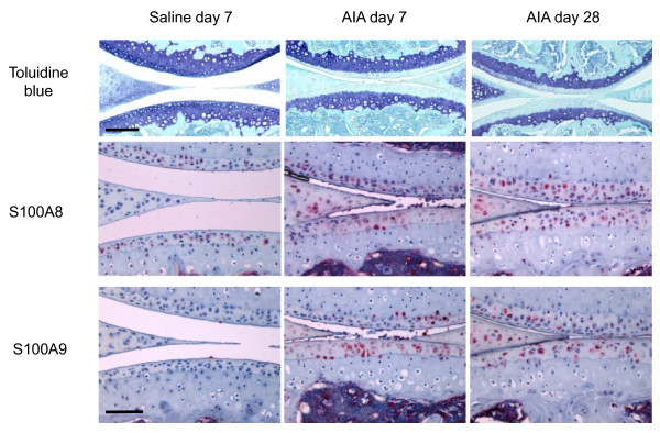
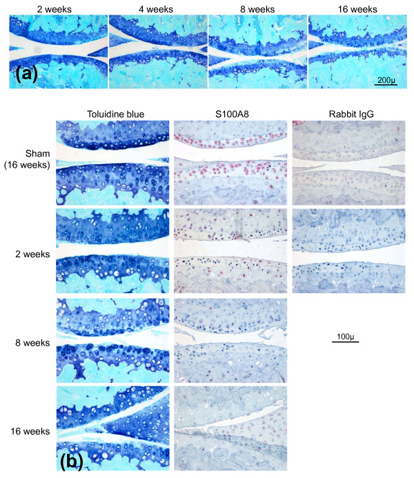
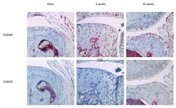
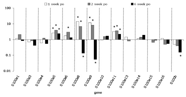
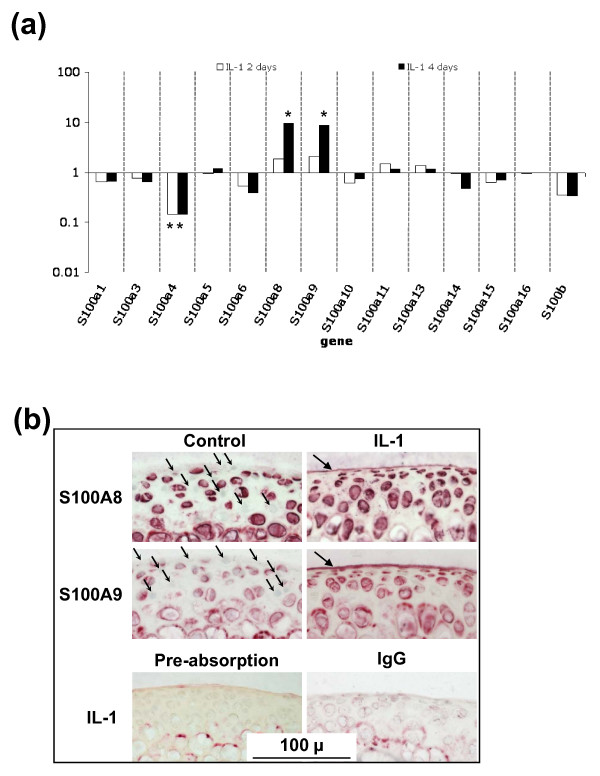
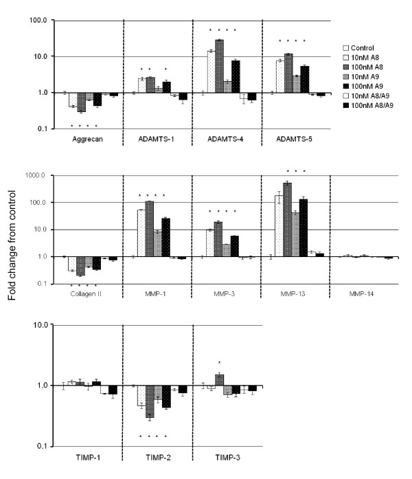
Methods: S100A8 and S100A9 protein localization were determined in antigen-induced inflammatory arthritis in mice, mouse femoral head cartilage explants stimulated with interleukin-1 (IL-1), and in surgically-induced OA in mice. Microarray expression profiling of all S100 proteins in cartilage was evaluated at different times after initiation of degradation in femoral head explant cultures stimulated with IL-1 and surgically-induced OA. The effect of S100A8, S100A9 or the complex on the expression of aggrecan (Acan), collagen II (Col2a1), disintegrin and metalloproteases with thrombospondin motifs (Adamts1, Adamts 4 &Adamts 5), matrix metalloproteases (Mmp1, Mmp3, Mmp13 &Mmp14) and tissue inhibitors of metalloproteinases (Timp1, Timp2 &Timp3), by primary adult ovine articular chondrocytes was determined using real time quantitative reverse transcription polymerase chain reaction (qRT-PCR).

Results: Stimulation with IL-1 increased chondrocyte S100a8 and S100a9 mRNA and protein levels. There was increased chondrocyte mRNA expression of S100a8 and S100a9 in early but not late mouse OA. However, loss of the S100A8 staining in chondrocytes occurred as mouse OA progressed, in contrast to the positive reactivity for both S100A8 and S100A9 in chondrocytes in inflammatory arthritis in mice. Homodimeric S100A8 and S100A9, but not the heterodimeric complex, significantly upregulated chondrocyte Adamts1, Adamts4 and Adamts 5, Mmp1, Mmp3 and Mmp13 gene expression, while collagen II and aggrecan mRNAs were significantly decreased.

Conclusions: Chondrocyte derived S100A8 and S100A9 may have a sustained role in cartilage degradation in inflammatory arthritis. In contrast, while these proteins may have a role in initiating early cartilage degradation in OA by upregulating MMPs and aggrecanases, their reduced expression in late stages of OA suggests they do not have an ongoing role in cartilage degradation in this non-inflammatory arthropathy.


Figures

Figure 1
Figure 1
Toluidine blue/fast green and S100A8 and S100A9 immunostained paraffin sections of medial femoro-tibial compartments of mouse knee joints from saline-injected compared with AIA at days 7 and 28. Scale bar = 100 μm. AIA = antigen-induced arthritis.
Figure 2
Figure 2
Histopathological examination of osteoarthritic changes in mouse knee (femoro-tibial) joints following medial meniscal destabilization (MMD). (a) Progressive cartilage damage at 2, 4, 8 and 16 weeks following medial meniscal destabilization-induced osteoarthritis (OA) in mouse knee joints. Toluidine blue/fast green stained paraffin sections. Scale bar = 200 μm. (b) Serial sections stained with toluidine blue or with S100A8 immunolocalization in mouse knees at different times following medial meniscal destabilization (MMD) or sham surgery (at 16 weeks). Scale bar = 100 μm. Negative control sections were immunostained using an equivalent concentration of rabbit IgG.
Figure 3
Figure 3
S100A8 and S100A9 immunostaining in cartilage and subchondral bone at the joint margins following sham operation or medial meniscal destabilization at 4 and 16 weeks. Scale bar = 100 μm.
Figure 4
Figure 4
Fold change in S100 gene expression measured by microarray expression profiling, in micro-dissected cartilage from surgically-induced OA compared with sham-operated joints 1, 2 and 6 weeks post-operatively. Pooled samples were used from three sham or medial meniscal destabilization (MMD) joints per time point. *B-statistic ≥ 1.0. OA = osteoarthritis; po = post-operatively.
Figure 5
Figure 5
Changes in S100 proteins in cultured mouse articular cartilage. (a) Fold change in S100 gene expression, measured by microarray expression profiling, in mouse femoral head cartilage cultured with or without IL-1 for two or four days compared with control cultures measured by microarray expression profiling (pooled sample from three femoral heads/treatment/time). *B-statistic ≥ 1.0. (b) S100A8 and S100A9 immunostaining in frozen sections of mouse femoral head articular cartilage following four days of culture with IL-1. Substantial numbers of chondrocytes did not stain with S100A8 and S100A9 in the control cultures (small arrows). In IL-1-stimulated cultures the intensity and proportion of positively stained chondrocytes increased. In addition, the surface matrix lamina stained positive for both S100A8 and S100A9 (large arrows). Negative controls included sections of IL-1-stimulated cartilage incubated with equal concentration of IgG, or localized with anti-S100A8 following pre-absorption with recombinant S100A8 protein. Scale bar = 100 μm.
Figure 6
Figure 6
Fold change (± SEM) compared with unstimulated cultures in gene expression measured by real-time qRT-PCR in primary ovine chondrocytes cultured for 24 hours in monolayer with 10 or 100 nM S100A8, S100A9 or S100A8/S100A9 complex. n = 6 per culture condition. * = significant difference compared with control cultures (P < 0.027).

Similar articles

Cited by

References

    1. Vogl T, Ludwig S, Goebeler M, Strey A, Thorey IS, Reichelt R, Foell D, Gerke V, Manitz MP, Nacken W, Werner S, Sorg C, Roth J. MRP8 and MRP14 control microtubule reorganization during transendothelial migration of phagocytes. Blood. 2004;104:4260–4268. doi: 10.1182/blood-2004-02-0446. - DOI - PubMed
    1. Cornish CJ, Devery JM, Poronnik P, Lackmann M, Cook DI, Geczy CL. S100 protein CP-10 stimulates myeloid cell chemotaxis without activation. J Cell Physiol. 1996;166:427–437. doi: 10.1002/(SICI)1097-4652(199602)166:2<427::AID-JCP21>3.0.CO;2-6. - DOI - PubMed
    1. Harrison CA, Raftery MJ, Walsh J, Alewood P, Iismaa SE, Thliveris S, Geczy CL. Oxidation regulates the inflammatory properties of the murine S100 protein S100A8. J Biol Chem. 1999;274:8561–8569. doi: 10.1074/jbc.274.13.8561. - DOI - PubMed
    1. Raftery MJ, Collinson L, Geczy CL. Overexpression, oxidative refolding, and zinc binding of recombinant forms of the murine S100 protein MRP14 (S100A9) Protein Expr Purif. 1999;15:228–235. doi: 10.1006/prep.1998.1015. - DOI - PubMed
    1. Cecil DL, Johnson K, Rediske J, Lotz M, Schmidt AM, Terkeltaub R. Inflammation-induced chondrocyte hypertrophy is driven by receptor for advanced glycation end products. J Immunol. 2005;175:8296–8302. - PubMed

Publication types

MeSH terms