. 2010 Jan 27:9:19.
doi: 10.1186/1476-4598-9-19.

The mitochondria-independent cytotoxic effect of nelfinavir on leukemia cells can be enhanced by sorafenib-mediated mcl-1 downregulation and mitochondrial membrane destabilization

Affiliations

The mitochondria-independent cytotoxic effect of nelfinavir on leukemia cells can be enhanced by sorafenib-mediated mcl-1 downregulation and mitochondrial membrane destabilization

Ansgar Brüning et al. Mol Cancer. .

Abstract

Background: Nelfinavir is an HIV protease inhibitor that has been used for a long period of time to treat HIV-infected individuals. It has recently emerged that nelfinavir could represent a prospective new anti-cancer drug, prompting us to test the effect of nelfinavir on leukemia cells.

Methods: By combining in vitro and ex vivo studies, the effect of nelfinavir on leukemia cells and non-malignant, bone marrow-derived tissue cells was analyzed.

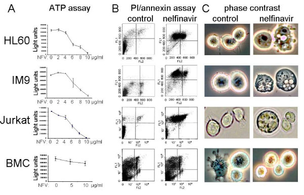
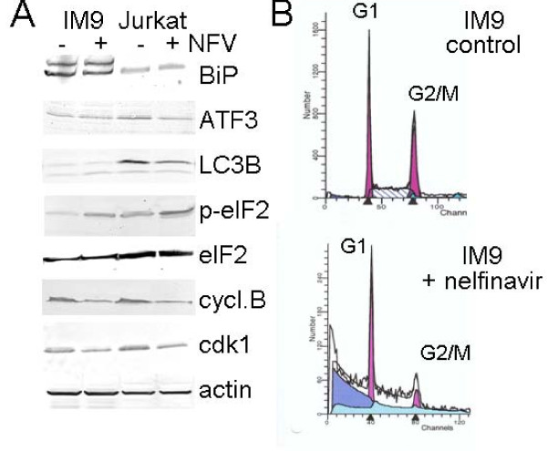
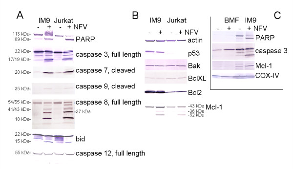
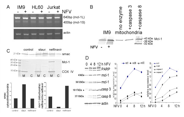
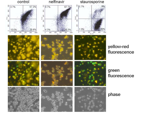
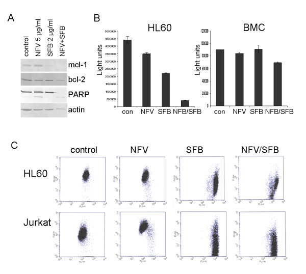
Results: At a concentration of 9 microg/ml, nelfinavir induced death of 90% of HL60, IM9, and Jurkat cells. At the same concentration and treatment conditions, less than 10% of aspirated human bone marrow cells showed nelfinavir-induced cell damage. Nelfinavir-induced death of leukemia cells was accompanied by activation of caspases 3, 7, and 8. Despite caspase activation, the upregulation of the anti-apoptotic bcl-2 family member protein mcl-1 that resulted from nelfinavir treatment stabilized the mitochondrial membrane potential, resulting in primarily mitochondria-independent cell death. Pharmacological downregulation of mcl-1 expression by treatment with sorafenib (2 microg/ml) significantly enhanced nelfinavir-induced apoptosis even at lower nelfinavir concentrations (5 microg/ml), but did not have additional detrimental effects on non-malignant bone marrow cells.

Conclusions: The ability of nelfinavir to induce apoptosis in leukemia cells as a single agent in a mitochondria-independent manner might suggest it could be used as a second or third line of treatment for leukemia patients for whom standard mitochondria-directed treatment strategies have failed. Combination treatment with nelfinavir and sorafenib might further enhance the efficacy of nelfinavir even on chemo-resistant leukemia cells.


Figures

Figure 1
Figure 1
Nelfinavir induces cell death in leukemia cells. The human leukemia cell lines HL60, Jurkat, and IM9, and an ex vivo sample of human bone marrow cells (BMC) were treated for 48 h in cell culture with the indicated concentrations of nelfinavir and analyzed by a chemiluminescent ATP assay for cell survival (A), by annexin/PI staining [FL-1 (propidium iodide): 575 nm; FL-2 (FITC): 530 nm] for the occurrence of apoptosis and necrosis (B), and by phase contrast microscopy for morphological changes (C). For FACScan analysis and phase contrast microscopy, all cells were treated equally for 24 h with 8 μg/ml nelfinavir.
Figure 2
Figure 2
Influence of nelfinavir on cell cycle progression and apoptosis in human leukemia cells. IM9 and Jurkat cells were treated for 24 h with 8 μg/ml nelfinavir and analyzed by (A) Western blot analysis for the expression of cell stress- and cell cycle-related proteins and by (B) FACScan analysis for cell cycle distribution of nelfinavir-treated IM9 cells.
Figure 3
Figure 3
Nelfinavir induces activation of caspase 3 and caspase 8 despite mcl-1 upregulation. IM9, Jurkat and bone marrow fibroblast cells (BMF) were treated for 24 h with 8 μg/ml nelfinavir and analyzed by Western blot analysis for the expression of caspases and apoptosis-relevant proteins. (A) and (B): cell lysates of IM9 and Jurkat cells; (C) cell lysates of IM9 and BMF.
Figure 4
Figure 4
Nelfinavir induces mitochondria protection despite caspase 8-mediated mcl-1 cleavage. A) The indicated leukemia cell lines were treated with 8 μg/ml nelfinavir for 24 h hours and subjected to PCR analysis to detect potential mcl-1 splice forms and transcriptional mcl-1 regulation. B) An enriched cellular mitochondria fraction of IM9 cells was treated with recombinant caspase 3 or caspase 8 as indicated and subjected to Western blot analysis for mcl-1 expression. Whole cell extracts of nelfinavir-treated IM9 cells were run in parallel for comparison. C) IM9 cells treated for 24 h with 8 μg/ml nelfinavir or 500 nM staurosporine were separated into a crude mitochondria fraction (M) and a crude cytosol fraction (C) and analyzed by Western blot for the distribution of mcl-1, smac and mitochondria-resident protein cytochrome c oxidase (COX IV). For direct comparison, mcl-1 and COX IV immunostaining were performed on the same blot. Staining intensities were analyzed with a BioRad gel documentation system using the BioRad Quantity One program (BioRad, Munich, Germany) and expressed as relative expression values. D) IM9 cells were treated with 10 μg/ml nelfinavir for the indicated times and analyzed by Western blot analysis. Selected bands (mcl1: 43, 36, 32 kDa; caspase 3: 17+19 kDa; caspase 8: 18 kDa) were densitometrically analyzed using the BioRad Quantity One program and plotted as relative expression values versus β-actin expression.
Figure 5
Figure 5
Nelfinavir-induced cell death is independent of mitochondrial membrane depolarization. IM9 cells were treated for 24 h with 8 μg/ml nelfinavir or 500 nM staurosporine and the outer mitochondrial membrane potential was analyzed using the MitoCapture kit (Alexis, Lörrach, Germany) by either FACScan analysis or fluorescence microscopy.
Figure 6
Figure 6
Sorafenib enhances the efficacy of nelfinavir-induced cell death by mcl-1 downregulation. A) HL60 cells were treated with 5 μg/ml nelfinavir or 2 μg/ml sorafenib either alone or in combination, and analyzed after 24 h for the expression of apoptosis-related proteins by Western blot analysis. B) HL60 cells and ex vivo bone marrow cells (BMC) were incubated using the same conditions as in (A) and analyzed after 48 h for total cell survival by an ATP assay. C) HL60 and Jurkat cells were treated with either 5 μg/ml nelfinavir or 2 μg/ml sorafenib either alone or in combination, and the outer mitochondrial membrane potential was analyzed after 24 h by FACScan analysis.

Similar articles

Cited by

References

    1. Gills JJ, Lopiccolo J, Tsurutani J, Shoemaker RH, Best CJ, Abu-Asab MS, Borojerdi J, Warfel NA, Gardner ER, Danish M, Hollander MC, Kawabata S, Tsokos M, Figg WD, Steeg PS, Dennis PA. Nelfinavir, A lead HIV protease inhibitor, is a broad-spectrum, anticancer agent that induces endoplasmic reticulum stress, autophagy, and apoptosis in vitro and in vivo. Clin Cancer Res. 2007;13:5183–5184. doi: 10.1158/1078-0432.CCR-07-0161. - DOI - PubMed
    1. Pyrko P, Kardosh A, Wang W, Xiong W, Schönthal AH, Chen TC. HIV-1 protease inhibitors nelfinavir and atazanavir induce malignant glioma death by triggering endoplasmic reticulum stress. Cancer Res. 2007;67:10920–10928. doi: 10.1158/0008-5472.CAN-07-0796. - DOI - PubMed
    1. Cohen J. HIV drug shows promise as potential cancer treatment. Science. 2007;317:1305. doi: 10.1126/science.317.5843.1305. - DOI - PubMed
    1. Plastaras JP, Vapiwala N, Ahmed MS, Gudonis D, Cerniglia GJ, Feldman MD, Frank I, Gupta AK. Validation and toxicity of PI3K/Akt pathway inhibition by HIV protease inhibitors in humans. Cancer Biol Ther. 2008;7:628–635. - PubMed
    1. Brunner TB, Geiger M, Grabenbauer GG, Lang-Welzenbach M, Mantoni TS, Cavallaro A, Sauer R, Hohenberger W, McKenna WG. Phase I trial of the human immunodeficiency virus protease inhibitor nelfinavir and chemoradiation for locally advanced pancreatic cancer. J Clin Oncol. 2008;26:2699–2706. doi: 10.1200/JCO.2007.15.2355. - DOI - PubMed

Publication types

MeSH terms