. 2010 Jan 27:8:7.
doi: 10.1186/1479-5876-8-7.

Boosting high-intensity focused ultrasound-induced anti-tumor immunity using a sparse-scan strategy that can more effectively promote dendritic cell maturation

Affiliations

Boosting high-intensity focused ultrasound-induced anti-tumor immunity using a sparse-scan strategy that can more effectively promote dendritic cell maturation

Fang Liu et al. J Transl Med. .

Abstract

Background: The conventional treatment protocol in high-intensity focused ultrasound (HIFU) therapy utilizes a dense-scan strategy to produce closely packed thermal lesions aiming at eradicating as much tumor mass as possible. However, this strategy is not most effective in terms of inducing a systemic anti-tumor immunity so that it cannot provide efficient micro-metastatic control and long-term tumor resistance. We have previously provided evidence that HIFU may enhance systemic anti-tumor immunity by in situ activation of dendritic cells (DCs) inside HIFU-treated tumor tissue. The present study was conducted to test the feasibility of a sparse-scan strategy to boost HIFU-induced anti-tumor immune response by more effectively promoting DC maturation.

Methods: An experimental HIFU system was set up to perform tumor ablation experiments in subcutaneous implanted MC-38 and B16 tumor with dense- or sparse-scan strategy to produce closely-packed or separated thermal lesions. DCs infiltration into HIFU-treated tumor tissues was detected by immunohistochemistry and flow cytometry. DCs maturation was evaluated by IL-12/IL-10 production and CD80/CD86 expression after co-culture with tumor cells treated with different HIFU. HIFU-induced anti-tumor immune response was evaluated by detecting growth-retarding effects on distant re-challenged tumor and tumor-specific IFN-gamma-secreting cells in HIFU-treated mice.

Results: HIFU exposure raised temperature up to 80 degrees centigrade at beam focus within 4 s in experimental tumors and led to formation of a well-defined thermal lesion. The infiltrated DCs were recruited to the periphery of lesion, where the peak temperature was only 55 degrees centigrade during HIFU exposure. Tumor cells heated to 55 degrees centigrade in 4-s HIFU exposure were more effective to stimulate co-cultured DCs to mature. Sparse-scan HIFU, which can reserve 55 degrees-heated tumor cells surrounding the separated lesions, elicited an enhanced anti-tumor immune response than dense-scan HIFU, while their suppressive effects on the treated primary tumor were maintained at the same level. Flow cytometry analysis showed that sparse-scan HIFU was more effective than dense-scan HIFU in enhancing DC infiltration into tumor tissues and promoting their maturation in situ.

Conclusion: Optimizing scan strategy is a feasible way to boost HIFU-induced anti-tumor immunity by more effectively promoting DC maturation.


Figures

Figure 1
Figure 1
The experimental HIFU system. (A) Diagram of the in vivo HIFU exposure setup. (B) A tumor-bearing mouse. (C) The way the mouse was fixed during HIFU exposure. (D) The B-mode ultrasound image of the tumor. (E) Diagram of the in vitro HIFU exposure setup.
Figure 2
Figure 2
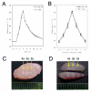
Thermal effects of HIFU treatment. (A) Temperature profiles at the beam focus in MC-38 and B16 tumors when the transducer was run in continuous wave (CW) mode at a pressure level of P+ = 19.5/P- = -7.2 MPa. Representative data of three independent experiments with consistent results are shown. (B) Lateral distribution of peak temperature in tumors produced by HIFU during 4-s exposures. Results are expressed as means ± SD out of four independent experiments. (C) Transversal and (D) longitudinal views of thermal lesions produced by HIFU with different treatment duration (4, 3, and 2 s) at above pressure level. The representative section from four treated mice with similar results is shown.
Figure 3
Figure 3
HIFU-induced DC infiltration surrounding the thermal lesion. Tumor tissue samples were collected 1 day after HIFU treatment. 6-μm cryostat sections were cut and stained with anti-CD11c Abs. Then the antibody was visualized using the Anti-Hamster Ig HRP detection kit. The sections were counterstained with hematoxylin. Representative sections from each group of four mice are shown.
Figure 4
Figure 4
DC maturation stimulated by HIFU-treated tumor cells. (A) Temperature profiles produced by 55°C-HIFU and 80°C-HIFU. (B) Immature DCs were incubated for 2 days in the presence of CpG-ODN, normal B16 cells, 55°C-HIFU and 80°C-HIFU treated B16 cells. Levels of IL-12 p70 and IL-10 in the culture supernatants were measured by ELISA. (C) Expression of CD80 and CD86 on the surface of DC (thick line) was assayed by Flow cytometry. Solid thin line represents the expression of these markers on surface of non-stimulated DC. Representative data out of three separate experiments are shown. (D) The expression levels of CD80 and CD86 on DCs were presented as mean fluorescence intensity. Results in panels B and D are expressed as means ± SD out of three independent experiments. * p < 0.05 compared with 'DC Alone', # p < 0.05 compared with 'DC+normal B16', ! p < 0.05 compared with 'DC+80°C-HIFU' by Student's t test.
Figure 5
Figure 5
Comparison of tumor ablation and systemic immune response induced by two different scan strategies. (A) Thermal lesions produced by dense- and sparse-scan strategies in MC-38 tumors. (B-C) The suppressive effects of different scan strategies on the growth of treated primary tumors. (D-E) The retarding effects on the growth of distant re-challenged tumors. (F-G) Tumor-specific IFN-γ-secreting cells detected in the splenocytes of HIFU-treated mice. C57BL/6 mice were inoculated s.c. on right hind leg with 5 × 10⁵ MC-38 or B16 tumor cells and treated with different HIFU on day 9 of tumor inoculation. Mice were challenged with 1 × 10⁶ MC-38 or B16 tumor cells by s.c. inoculation on the left hind leg one day after HIFU treatment. Both primary and challenged tumor growth was monitored daily. Tumor-specific IFN-γ-secreting cells were detected in splenocytes by ELISPOTS assays. Results were expressed as mean ± SD for each group (n = 8 per group). *P < 0.05; **P < 0.001 versus non-treatment control by Student's t test. This experiment is representative of three experiments with consistent results.
Figure 6
Figure 6
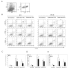
DCs were recruited into tumor tissues one day after HIFU treatment and exhibited the surface phenotype of maturation. (A) The presence of CD45+ tumor-infiltrating leukocytes in tumor tissues was identified in the gate indicated. (B) CD11c+ cells in the gate defined in A were analyzed for the expression of MHC II, CD80, and CD86. Representative data of six independent experiments with consistent results are shown. (C) The proportion of tumor-infiltrating DC (CD11c+/MHC II+) (expressed in percentage of total cells) was investigated for the indicated tumors one day after different HIFU-treatment. (D) The expression levels of CD86 (presented as mean fluorescence intensity) were analyzed in CD11c+ cells infiltrating B16 or MC-38 tumor one day after different HIFU-treatment. (E) The expression levels of CD80 (presented as mean fluorescence intensity) were analyzed in CD11c+ cells infiltrating B16 or MC-38 tumor one day after different HIFU-treatment. (C-E) Results were expressed as mean ± SD for each group (n = 6 per group). *P < 0.05 versus non-treatment control; #P < 0.05 versus Dense-scan HIFU by Student's t test. This experiment is representative of three experiments with consistent results.

References

    1. Hynynen K, Pomeroy O, Smith DN, Huber PE, McDannold NJ, Kettenbach J, Baum J, Singer S, Jolesz FA. MR imaging-guided focused ultrasound surgery of fibroadenomas in the breast: a feasibility study. Radiology. 2001;219:176–185. - PubMed
    1. Chaussy C, Thuroff S. The status of high-intensity focused ultrasound in the treatment of localized prostate cancer and the impact of a combined resection. Curr Urol Rep. 2003;4:248–252. doi: 10.1007/s11934-003-0077-0. - DOI - PubMed
    1. Kennedy JE, Wu F, ter Haar GR, Gleeson FV, Phillips RR, Middleton MR, Cranston D. High-intensity focused ultrasound for the treatment of liver tumours. Ultrasonics. 2004;42:931–935. doi: 10.1016/j.ultras.2004.01.089. - DOI - PubMed
    1. Chen W, Wang Z, Wu F, Zhu H, Zou J, Bai J, Li K, Xie F. [High intensity focused ultrasound in the treatment of primary malignant bone tumor] Zhonghua Zhong Liu Za Zhi. 2002;24:612–615. - PubMed
    1. Zhang L, Chen WZ, Liu YJ, Hu X, Zhou K, Chen L, Peng S, Zhu H, Zou HL, Bai J, Wang ZB. Feasibility of magnetic resonance imaging-guided high intensity focused ultrasound therapy for ablating uterine fibroids in patients with bowel lies anterior to uterus. Eur J Radiol. 2008. - PubMed

Publication types

LinkOut - more resources