

Hepatic pulmonary fusion in an infant with a right-sided congenital diaphragmatic hernia and contralateral mediastinal shift
- PMID: 20105618
- PMCID: PMC4418537
- DOI: 10.1016/j.jpedsurg.2009.10.090
Hepatic pulmonary fusion in an infant with a right-sided congenital diaphragmatic hernia and contralateral mediastinal shift
Abstract
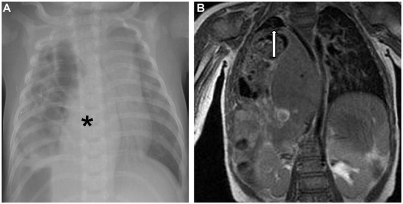
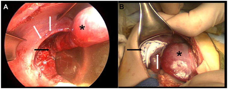
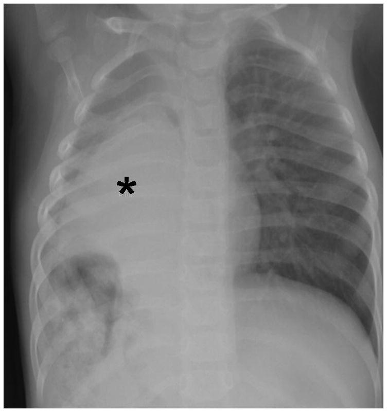
Hepatic pulmonary fusion is extremely rare with only 9 previous cases reported in the literature. In typical cases, the clinician should be alerted to the possibility of hepatic pulmonary fusion if the chest radiograph shows a large opacity on the right side without a contralateral mediastinal shift. The authors present a case of right-sided diaphragmatic hernia and hepatic pulmonary fusion with associated contralateral mediastinal shift discovered beyond the neonatal period. The 9 previous cases were retrospectively reviewed with special attention to mediastinal shift on preoperative chest radiograph, operative procedure, and mortality. Only one previous case demonstrated a contralateral mediastinal shift. The most common procedure performed was partial separation of the hepatic pulmonary fusion and approximation of the diaphragmatic defect. Four of the previous 9 patients died. In our case, reduction of bowel and approximation of the diaphragmatic defect around the fused liver and lung have been successful.
Copyright 2010 Elsevier Inc. All rights reserved.
Figures
References
-
- Fisher JC, Jefferson RA, Arkovitz MS, et al. Redefining outcomes in right congenital diaphragmatic hernia. J Pediatr Surg. 2008;43:373–9. - PubMed
-
- Weber TR, Tracy T, Bailey PV, et al. Congenital diaphragmatic hernia beyond infancy. Am J Surg. 1991;162:643–6. - PubMed
-
- Robertson DJ, Harmon CM, Goldberg S. Right congenital diaphragmatic hernia associated with fusion of the liver and the lung. J Pediatr Surg. 2006;41:9–10. - PubMed
-
- Slovis TL, Farmer DL, Berdon WE, et al. Hepatic Pulmonary Fusion in Neonates. AJR. 2000;174:229–33. - PubMed
-
- Keller RL, Aaroz PA, Hawgood S, et al. MR Imaging of Hepatic Pulmonary Fusion in Neonates. AJR. 2003;180:438–40. - PubMed
Publication types
MeSH terms
Grants and funding
LinkOut - more resources
Full Text Sources
