Review
. 2010 May 7;277(1686):1299-307.
doi: 10.1098/rspb.2009.2195. Epub 2010 Jan 27.

Resolving phylogenetic incongruence to articulate homology and phenotypic evolution: a case study from Nematoda

Affiliations
Review

Resolving phylogenetic incongruence to articulate homology and phenotypic evolution: a case study from Nematoda

Erik J Ragsdale et al. Proc Biol Sci. .

Abstract

Modern morphology-based systematics, including questions of incongruence with molecular data, emphasizes analysis over similarity criteria to assess homology. Yet detailed examination of a few key characters, using new tools and processes such as computerized, three-dimensional ultrastructural reconstruction of cell complexes, can resolve apparent incongruence by re-examining primary homologies. In nematodes of Tylenchomorpha, a parasitic feeding phenotype is thus reconciled with immediate free-living outgroups. Closer inspection of morphology reveals phenotypes congruent with molecular-based phylogeny and points to a new locus of homology in mouthparts. In nematode models, the study of individually homologous cells reveals a conserved modality of evolution among dissimilar feeding apparati adapted to divergent lifestyles. Conservatism of cellular components, consistent with that of other body systems, allows meaningful comparative morphology in difficult groups of microscopic organisms. The advent of phylogenomics is synergistic with morphology in systematics, providing an honest test of homology in the evolution of phenotype.


Figures

Figure 1.
Figure 1.
The epidermis, muscle and cuticle of the anterior end of three representatives of Rhabditida: (a) A. complexus, (b) C. elegans and (c) A. avenae. Stoma/stomatostylet characters understood from three-dimensional TEM reconstruction (coloured diagrams/models) are contrasted with morphology seen by differential interference contrast (DIC) light microscopy (right of each diagram). In spite of gross divergence in feeding structures noted by several generations of taxonomists, closer examination of morphology reveals that underlying syncytia are highly conserved in number and in spatial relationships (in addition to numbers and positions of cell bodies, all not shown), allowing reliable assessments of homology. Corresponding colours represent putative homologues. The stoma lining that is falsely coloured darker grey is putatively homologous in all three taxa and is underlain, and presumably produced (Wright & Thomson 1981; Endo 1985), by the arcade syncytia and ‘e1’ in all cases, but it is expressed as a stomatostylet in the tylenchid and as an open stoma in microbivores. Epithelium ‘HypD’ between Hyp3 and Hyp4 is known only in nematodes of putative sister groups Cephalobomorpha and Tylenchomorpha; e1 is expressed as an epithelial collar cell (not muscle) only in the rhabditid. (d) Model of stomatostylet and associated cells/syncytia of A. avenae, showing nuances of cell morphology captured by three-dimensional TEM reconstruction, corresponding to the diagram and DIC image. The model is reconstructed directly from data presented in Ragsdale et al. (2008); the arcade syncytia and stomatostylet are shown complete; only one (dorsal) e1 muscle is shown; all other cuticle and syncytia are cut away transversely. Diagrammatic representations of stoma and associated cells/syncytia are informed by TEM reconstructions by Bumbarger et al. (2006) for A. complexus, by Wright & Thomson (1981), White (1988), De Ley et al. (1995) and Hall & Altun (2008) for C. elegans, and by Ragsdale (2008, 2009) for A. avenae. Abbreviations: Hyp, epidermis cell/syncytium; e1, e1 muscle/epithelial cell. Red, posterior arcade; orange, anterior arcade; yellow, Hyp1; green, Hyp2; light blue, Hyp3; dark blue, HypD; violet, Hyp4; brown, e1; light grey, cuticle; dark grey, stoma lining.
Figure 2.
Figure 2.
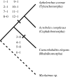
Simplified relationships between nematode models with reconstructed sensory anatomies, with preliminary mapping of characters. Ingroup relationships are a consensus of published phylogenies derived from 18S and 28S rRNA (Holterman et al. 2006; Nadler et al. 2006; Smythe et al. 2006; Meldal et al. 2007; van Megen et al. 2009). Broken line indicates a probable position of Myolaimus sp. based on ongoing morphological and molecular studies (De Ley & Blaxter 2002; Nadler et al. 2006). All extant studies are unanimous in placing Myolaimus sp. on a branch between cephalobids + tylenchids and representative rhabditids + diplogasterids, consistent with the single state changes shown for all characters in table 1. Characters are mapped under simple parsimony. Characters with missing states in Myolaimus sp. are mapped as parsimonious gains, pending further outgroup analysis. Numbers of characters (left of dash) and states (right of dash) correspond to characters drawn from table 1.

Similar articles

Cited by

References

    1. Albertson D. G., Thomson J. N.1976The pharynx of Caenorhabditis elegans. Phil. Trans. R. Soc. Lond. B 275, 299–325 (doi:10.1098/rstb.1976.0085) - DOI - PubMed
    1. Aleshin V. V., Kedrova O. S., Milyutina I. A., Vladychenskaya N. S., Petrov N. B.1998Relationships among nematodes based on the analysis of 18S rRNA gene sequences: molecular evidence for monophyly of chromadorian and secernentean nematodes. Russ. J. Nematol. 6, 175–184
    1. Andrássy I.1962Über den Mundstachel der Tylenchiden (Nematologische Notizen, 9). Acta. Zool. Hung. 8, 167–315
    1. Andrássy I.1976Evolution as a basis for the systemization of nematodes London, UK: Pitman Publishing
    1. Ashton F. T., Bhopale V. M., Fine A. E., Schad G. A.1995Sensory neuroanatomy of a skin-penetrating nematode parasite: Strongyloides stercoralis. I. Amphidial neurons. J. Comp. Neurol. 357, 281–295 (doi:10.1002/cne.903570208) - DOI - PubMed

Publication types

LinkOut - more resources