. 2010 Apr 15;19(8):1603-17.
doi: 10.1093/hmg/ddq036. Epub 2010 Jan 27.

Genetic studies on the functional relevance of the protein prenyltransferases in skin keratinocytes

Affiliations

Genetic studies on the functional relevance of the protein prenyltransferases in skin keratinocytes

Roger Lee et al. Hum Mol Genet. .

Abstract

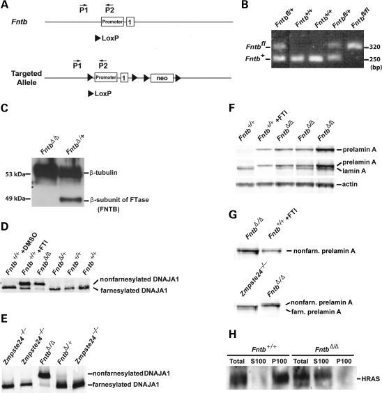
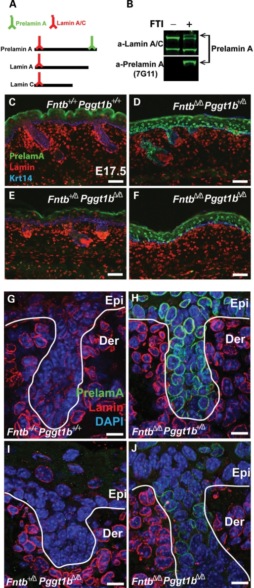
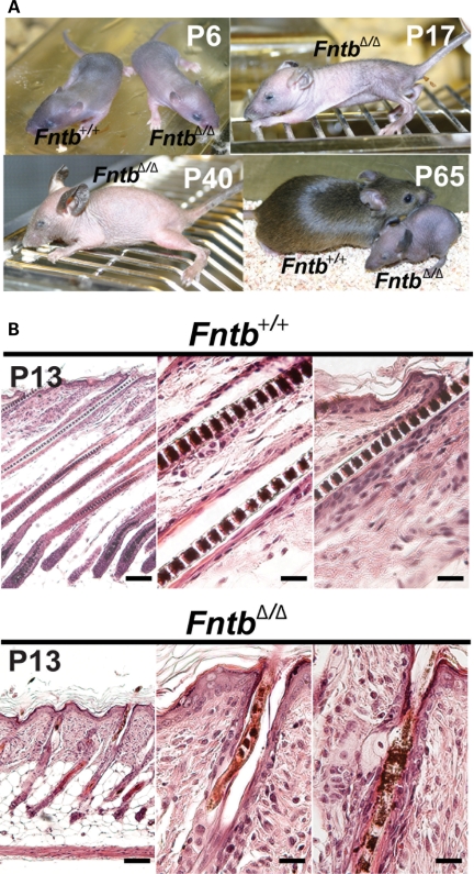
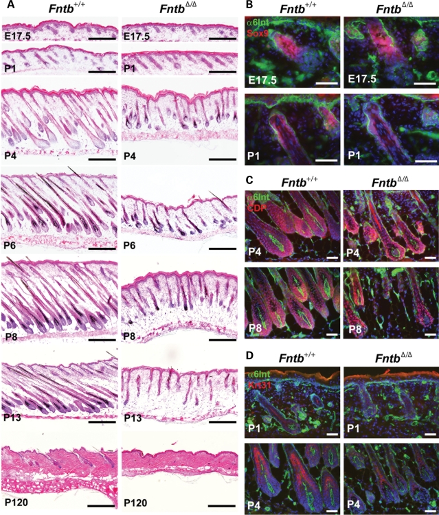
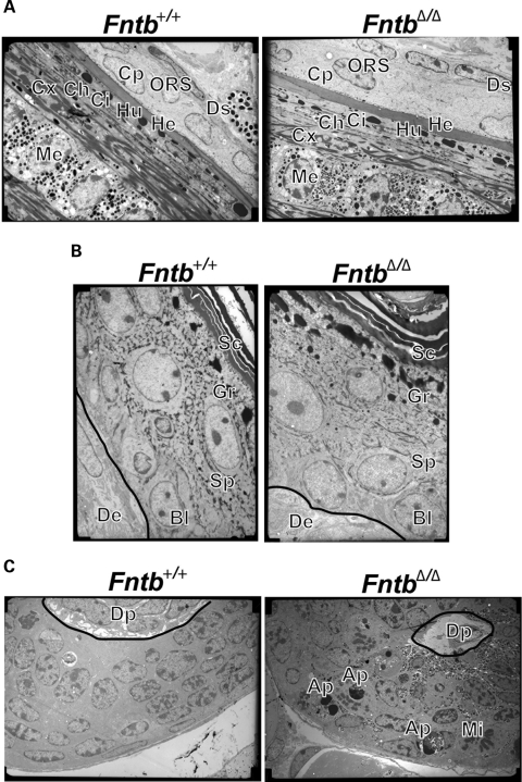
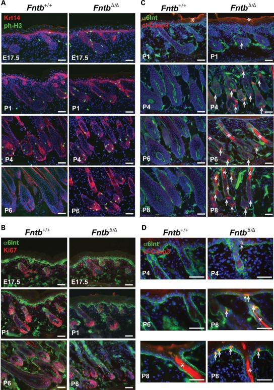
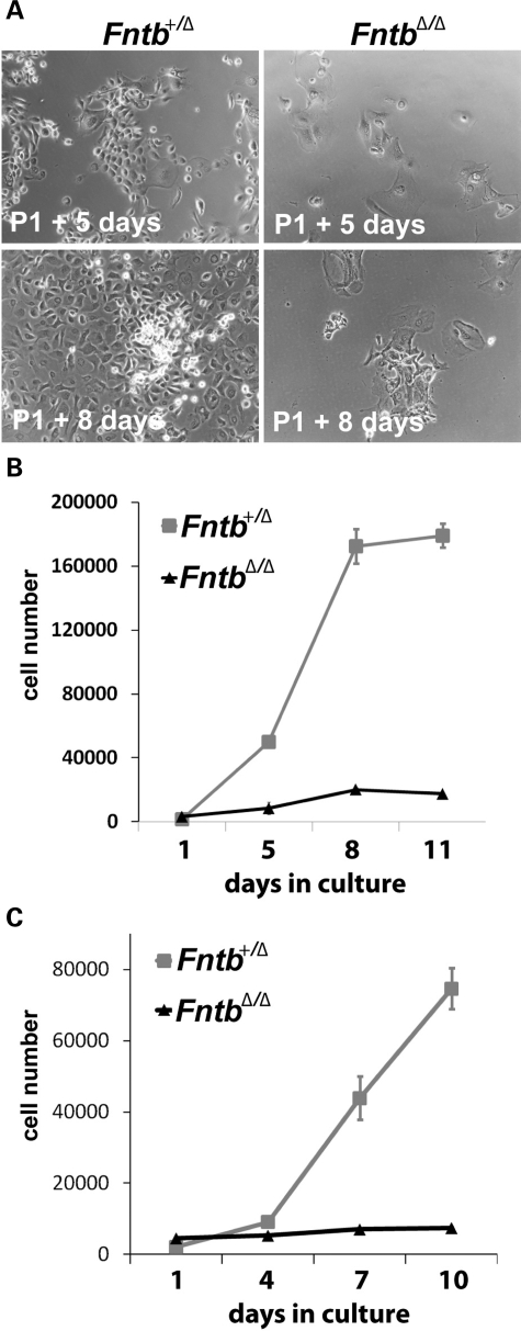
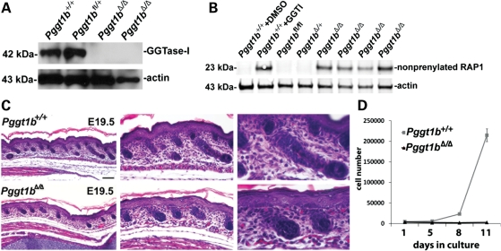
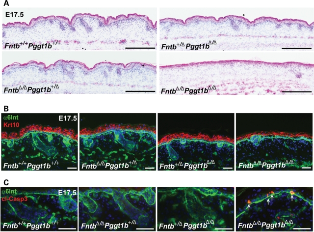
The modification of proteins with farnesyl or geranylgeranyl lipids, a process called protein prenylation, facilitates interactions of proteins with membrane surfaces. Protein prenylation is carried out by a pair of cytosolic enzymes, protein farnesyltransferase (FTase) and protein geranylgeranyltransferase type I (GGTase-I). FTase and GGTase-I have attracted interest as therapeutic targets for both cancer and progeria, but very little information exists on the importance of these enzymes for homeostasis of normal tissues. One study actually suggested that FTase is entirely dispensable. To explore the importance of the protein prenyltransferases for normal tissues, we used conditional knockout alleles for Fntb and Pggt1b (which encode the beta-subunits of FTase and GGTase-I, respectively) and a keratin 14-Cre transgene to create mice lacking FTase or GGTase-I in skin keratinocytes. Keratinocyte-specific Fntb knockout mice were viable but developed severe alopecia. Although hair follicles appeared normal during development, they were morphologically abnormal after birth, and ultrastructural and immunohistochemical studies revealed many apoptotic cells. The interfollicular epidermis of Fntb-deficient mice appeared normal; however, keratinocytes from these mice could not proliferate in culture. As expected, non-farnesylated prelamin A and non-farnesylated DNAJA1 accumulated in Fntb-deficient keratinocytes. Keratinocyte-specific Pggt1b knockout mice survived development but died shortly after birth. Like Fntb-deficient keratinocytes, Pggt1b-deficient keratinocytes did not proliferate in culture. Thus, both FTase and GGTase-I are required for the homeostasis of skin keratinocytes.


Figures

Figure 1.
Figure 1.
Inactivating FTase in keratinocytes. (A) Wild-type and conditional knockout alleles for Fntb (Fntb+ and Fntbfl, respectively). LoxP sites, indicated by a triangle, are located upstream of the Fntb promoter and in intron 1. (B) PCR-amplified fragments of Fntb+ and Fntbfl alleles, generated with the indicated primers, could be resolved on an ethidium bromide-stained 1.5% agarose gel. (C) Western blot of wild-type and FntbΔ/Δ keratinocytes with an antibody against the β-subunit of FTase (FNTB); β-tubulin was used as a loading control. (D, E) Western blot of keratinocyte extracts with a DNAJA1-specific monoclonal antibody, revealing that most of the DNAJA1 in FntbΔ/Δ keratinocytes was non-farnesylated. As a control, we examined DNAJA1 in cells that had been treated with an FTI (ABT-100, 5 μm). (F) Western blot of keratinocyte extracts with antibodies against prelamin A (top panel) or mature lamin A (the mature lamin A antibody binds both prelamin A and mature lamin A) (middle panel). Actin was used as a loading control. (G) Western blot with a prelamin A-specific polyclonal antibody showing that the prelamin A in FntbΔ/Δ keratinocytes co-migrates with non-farnesylated prelamin A in FTI-treated cells and migrates slightly more slowly than farnesylated prelamin A in Zmpste24−/− fibroblasts (56). (H) Western blots of HRAS in total cell extracts and P100 and S100 fractions of wild-type and FntbΔ/Δ keratinocytes.
Figure 2.
Figure 2.
Experiments with a new rat monoclonal antibody against prelamin A, 7G11, showing that prelamin A accumulates in FntbΔ/Δ keratinocytes. (A) Schematic depicting prelamin A, lamin A and lamin C and their recognition by the indicated antibodies. (B) Western blot of wild-type fibroblasts in the presence and absence of an FTI (ABT-100, 5 μm), showing that antibody 7G11 binds avidly to non-farnesylated prelamin A (lower panel). The detection of lamin A, lamin C and prelamin A with an N-terminal lamin A/C antibody is shown in the upper panel. (CJ) Immunohistochemical staining of skin with the prelamin A-specific antibody 7G11, a polyclonal antibody against lamins A/C and an antibody against keratin 14 at embryonic day 17.5. (C, G) Skin from a wild-type mouse (Fntb+/+Pggt1b+/+). (D, H) Skin from an FntbΔ/ΔPggt1b+/Δ mouse. (E, I) Skin from an Fntb+/ΔPggt1bΔ/Δ mouse. (F, J) Skin from an FntbΔ/ΔPggt1bΔ/Δ mouse. Scale bars for (C–F) are 50 µm and for (G–J) 16 µm.
Figure 3.
Figure 3.
Alopecia in keratinocyte-specific Fntb knockout mice (FntbΔ/Δ). (A) Alopecia in FntbΔ/Δ mice was detectable by close visual inspection on post-natal day 6 (P6) but was obvious at later time points (P17, P40, P65). (B) Hematoxylin and eosin-stained sections of skin from Fntb+/+ and FntbΔ/Δ mice at P13. Hair follicles in FntbΔ/Δ mice were stunted compared with those of Fntb+/+ mice, and the morphology of hair shafts was abnormal. Scale bars indicate 100 µm for left panels and 25 µm for middle and right panels.
Figure 4.
Figure 4.
Analysis of alopecia in keratinocyte-specific Fntb knockout mice (FntbΔ/Δ). (A) Hematoxylin and eosin-stained sections of skin from Fntb+/+ and FntbΔ/Δ mice at different time points. At P120, few hair follicles remained in FntbΔ/Δ mice. (B) Immunohistochemical staining of skin from Fntb+/+ and FntbΔ/Δ mice with an antibody against the stem cell marker Sox9 (red) indicates that the stem cell compartment is relatively unaffected by FTase deficiency. An antibody against α6-integrin (α6-Int, green) was used to demark epidermal structures. (C) Immunohistochemical staining of skin from Fntb+/+ and FntbΔ/Δ mice with an antibody against CDP (red), a marker of the follicular transit-amplifying population shows that the matrix of the hair follicle is present, even in the absence of FTase. However, the hair matrix was smaller in FntbΔ/Δ mice. (D) Immunohistochemical staining of skin from Fntb+/+ and FntbΔ/Δ mice with an antibody against keratin 31 (red), which is expressed in differentiating cells of the hair follicle. Keratin 31 was present in FntbΔ/Δ hair follicles, but fewer positive cells were detected. Scale bars indicate 50 µm for (B–D) and 200 µm for (A).
Figure 5.
Figure 5.
Electron microscopy of skin in Fntb+/+ and FntbΔ/Δ mice. (A) Electron micrographs of hair shafts, revealing that all layers of the hair shaft are present in FntbΔ/Δ mice, but the hair shafts were smaller than those in Fntb+/+ mice. Each layer of the follicle is demarcated: Ds, dermal sheath; ORS, outer root sheath; Cp, companion layer; He, Henley's layer; Hu, Huxley's layer; Ch and Ci, cuticle layers; Cx, cortex; Me, medulla. (B) Electron micrographs, showing that all layers of the interfollicular epidermis are intact in FntbΔ/Δ mice. De, dermis; Bl, basal layer; Sp, spinous layer; Gr, granular layer; Sc, stratum corneum. (C) Electron micrographs of the hair follicle matrix in Fntb+/+ and FntbΔ/Δ mice, revealing multiple apoptotic epithelial cells (Ap) and mitotic cells (Mi) in the matrix of the hair follicle (Mi) in FntbΔ/Δ mice. Images were acquired at 2900×.
Figure 6.
Figure 6.
Fntb deficiency in the epidermis does not affect proliferation in hair follicles but leads to increased numbers of apoptotic cells. (A) Immunohistochemical staining of skin with an antibody against phosphorylated histone-H3 (ph-H3, green), a marker of cell proliferation, reveals similar levels of staining in Fntb+/+ and FntbΔ/Δ mice. Keratin 14 staining (red) was used to demark epidermal cells. (B) Immunohistochemical studies with another marker of cell proliferation, Ki67, revealing many dividing cells in the skin from both Fntb+/+ and FntbΔ/Δ mice. (C) Immunohistochemical studies with an antibody against the cleaved form of caspase 3 (cl-Casp3, red), a marker of apoptotic cells, uncovered apoptotic cells in the hair follicles of FntbΔ/Δ mice (arrows) but not in Fntb+/+ mice. Hair shafts have a high level of autofluorescence (asterisks). (D) Additional immunohistochemical studies, focussing on the interfollicular epidermis, with an antibody against the cleaved form of caspase 3 (red). Apoptotic cells (arrows) could be identified in the interfollicular epidermis of FntbΔ/Δ mice. Scale bars indicate 50 µm for (A–D).
Figure 7.
Figure 7.
Fntb deficiency abolishes the ability of keratinocytes to proliferate. (A) Phase contrast images of keratinocytes from P1 Fntb+/Δ and FntbΔ/Δ mice after 5 or 8 days in culture. (B, C) Quantitative analysis of keratinocyte growth. Equal numbers of keratinocytes from Fntb+/Δ and FntbΔ/Δ mice at P1 (B) and P4 (C) were plated on 24-well plates. At the indicated time points, cells (3 wells/genotype) were counted. Data are representative of three independent experiments.
Figure 8.
Figure 8.
Inactivating GGTase-I in skin keratinocytes. (A) Western blot of keratinocytes from Pggt1b+/+and Pggt1bΔ/Δ mice with an antibody against the β-subunit of GGTase-I. Actin was used as a loading control. (B) Western blot of keratinocyte extracts with an antibody that recognizes the non-prenylated form of RAP1. As a positive control, we used Pggt1b+/+cells that had been treated with an inhibitor of GGTase-I (GGTI-298, 10 µm). (C) Hematoxylin and eosin-stained sections of skin from wild-type and Pggt1bΔ/Δ keratinocytes at E19.5, revealing stunted hair follicle growth in Pggt1bΔ/Δ mice. The interfollicular epidermis appeared normal (same image is shown at 10×, 20× and 40×). (D) Quantitative analysis of keratinocyte growth in culture. Equal numbers of keratinocytes from Pggt1b+/+ and Pggt1bΔ/Δ mice at E19.5 were plated on 24-well dishes, and cell proliferation was monitored for 10 days.
Figure 9.
Figure 9.
Inactivating both FTase and GGTase-I results in fewer hair follicles and a thinner interfollicular epidermis at E17.5. (A) Hematoxylin and eosin-stained sections of skin from Fntb+/+Pggt1b+/+, Fntb+/ΔPggt1bΔ/Δ, FntbΔ/ΔPggt1b+/Δ and FntbΔ/ΔPggt1bΔ/Δ embryos at E17.5. Hair follicle development in FntbΔ/Δ Pggt1bΔ/Δ embryos was stunted, and the skin was thinner, compared with mice lacking only FTase or only GGTase-I. (B) Staining for the spinous layer of the epidermis with an antibody against keratin 10 (Krt10, red) highlighted the thinning of the interfollicular epidermis in FntbΔ/ΔPggt1bΔ/Δ embryos. (C) Immunohistochemical studies with an antibody against the cleaved form of caspase 3 (cl-Casp3, red), revealing apoptotic epidermal cells in E17.5 FntbΔ/ΔPggt1bΔ/Δ epidermis (arrows). Scale bars indicate 50 µm for (B and C) and 500 µm for (A).

Similar articles

Cited by

References

    1. Casey P.J., Seabra M.C. Protein prenyltransferases. J. Biol. Chem. 1996;271:5289–5292. - PubMed
    1. Zhang F.L., Casey P.J. Protein prenylation: molecular mechanisms and functional consequences. Annu. Rev. Biochem. 1996;65:241–269. - PubMed
    1. Boyartchuk V.L., Ashby M.N., Rine J. Modulation of Ras and a-factor function by carboxyl-terminal proteolysis. Science. 1997;275:1796–1800. - PubMed
    1. Kim E., Ambroziak P., Otto J.C., Taylor B., Ashby M., Shannon K., Casey P.J., Young S.G. Disruption of the mouse Rce1 gene results in defective Ras processing and mislocalization of Ras within cells. J. Biol. Chem. 1999;274:8383–8390. - PubMed
    1. Otto J.C., Kim E., Young S.G., Casey P.J. Cloning and characterization of a mammalian prenyl protein-specific protease. J. Biol. Chem. 1999;274:8379–8382. - PubMed

Publication types

Substances