. 2010 May 1;19(9):1633-50.
doi: 10.1093/hmg/ddq038. Epub 2010 Jan 27.

Extensive enteric nervous system abnormalities in mice transgenic for artificial chromosomes containing Parkinson disease-associated alpha-synuclein gene mutations precede central nervous system changes

Affiliations

Extensive enteric nervous system abnormalities in mice transgenic for artificial chromosomes containing Parkinson disease-associated alpha-synuclein gene mutations precede central nervous system changes

Yien-Ming Kuo et al. Hum Mol Genet. .

Abstract

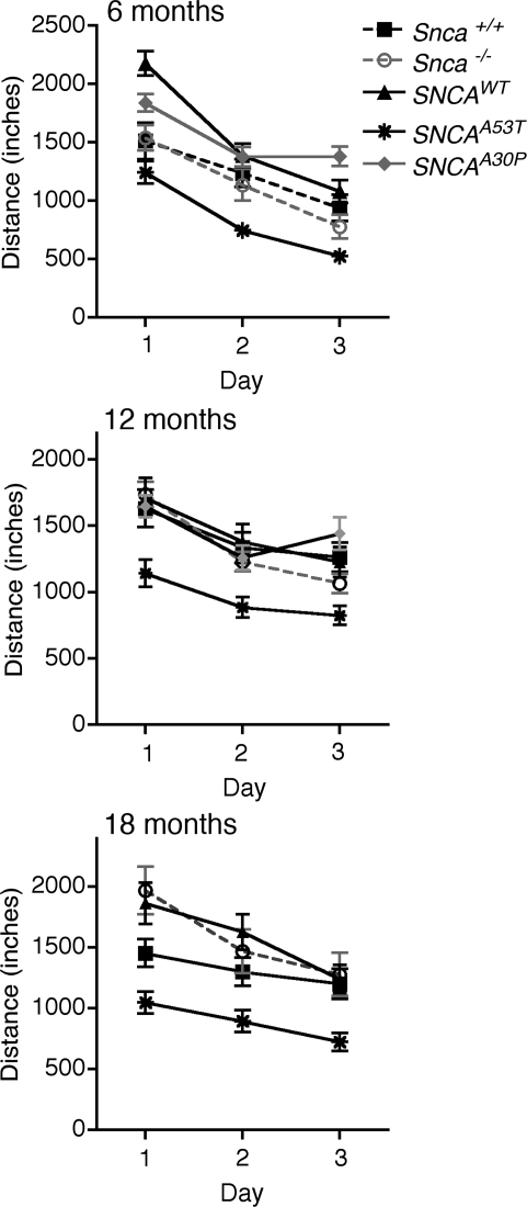
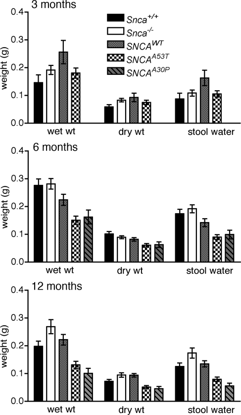
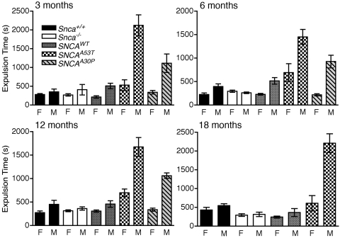
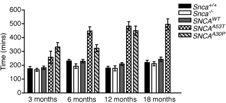
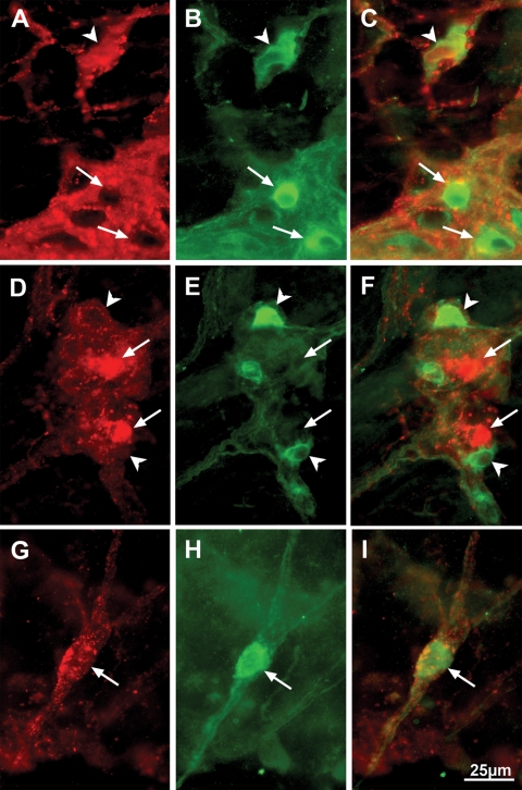
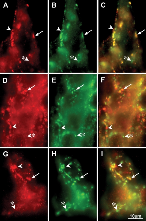
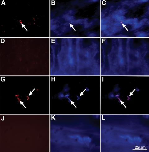
Parkinson disease (PD) is a neurodegenerative disease with motor as well as non-motor signs in the gastrointestinal tract that include dysphagia, gastroparesis, prolonged gastrointestinal transit time, constipation and difficulty with defecation. The gastrointestinal dysfunction commonly precedes the motor symptoms by decades. Most PD is sporadic and of unknown etiology, but a fraction is familial. Among familial forms of PD, a small fraction is caused by missense (A53T, A30P and E46K) and copy number mutations in SNCA which encodes alpha-synuclein, a primary protein constituent of Lewy bodies, the pathognomonic protein aggregates found in neurons in PD. We set out to develop transgenic mice expressing mutant alpha-synuclein (either A53T or A30P) from insertions of an entire human SNCA gene as models for the familial disease. Both the A53T and A30P lines show robust abnormalities in enteric nervous system (ENS) function and synuclein-immunoreactive aggregates in ENS ganglia by 3 months of age. The A53T line also has abnormal motor behavior but neither demonstrates cardiac autonomic abnormalities, olfactory dysfunction, dopaminergic neurotransmitter deficits, Lewy body inclusions or neurodegeneration. These animals recapitulate the early gastrointestinal abnormalities seen in human PD. The animals also serve as an in vivo system in which to investigate therapies for reversing the neurological dysfunction that target alpha-synuclein toxicity at its earliest stages.


Figures

Figure 1.
Figure 1.
Transcript and protein expression in brain and distal colon. (A) qPCR measurements in 6-week-old mice of human SNCA transcript relative to endogenous mouse Snca, using the housekeeping genes Elavl4 or synaptophysin as reference. Abbreviations are: SNCAWT is PAC-Tg(SNCAWT)+/+;Snca−/−, SNCAA53T is Dbl-PAC-Tg(SNCAA53T)+/+;Snca−/− and SNCAA30P is Dbl-PAC-Tg(SNCAA30P)+/+;Snca−/−, Elav is Elavl4, Syp is synpatophysin. Transcript levels relative to endogenous Snca are shown on a logarithmic scale. Each data point represents the average of four replicates per sample averaged over 10 mice of the same genotype. qPCR data and calculations are in Supplementary Material, Table S1A and B. (B) Western blot of α-synuclein protein in 6-month-old mice with either endogenous α-tubulin (brain) or β-actin (colon) as loading controls. Lane 1, Snca−/−; Lane 2, Snca+/+; Lane 3, PAC-Tg(SNCAWT)+/+;Snca−/−; Lane 4, Dbl-PAC-Tg(SNCAA53T)+/+;Snca−/−; Lane 5, Dbl-PAC-Tg(SNCAA30P)+/+;Snca−/−.
Figure 2.
Figure 2.
Latency (time to fall) in the Rotarod test at 6, 12 and 18 months of age. The pattern code corresponding to the various genotypes is given in the legend. Abbreviations are: SNCAWT is PAC-Tg(SNCAWT)+/+;Snca−/− and SNCAA53T is Dbl-PAC-Tg(SNCAA53T)+/+;Snca−/−. Error bars are ±1 standard errors of the mean. The number of mice of each genotype tested at each age is given in Supplementary Material, Table S2.
Figure 3.
Figure 3.
Total distance travelled in open field at 6, 12 and 18 months of age. Symbols distinguishing the line graphs for the different genotypes are in the legend. There are no data at the 18 month time point for the Dbl-PAC-Tg(SNCAA30P)+/+;Snca−/− line. Abbreviations are: SNCAWT is PAC-Tg(SNCAWT)+/+;Snca−/−, SNCAA53T is Dbl-PAC-Tg(SNCAA53T)+/+;Snca−/− and SNCAA30P is Dbl-PAC-Tg(SNCAA30P)+/+;Snca−/−. Error bars are ±1 standard errors of the mean. The number of mice of each genotype tested at each age is given in Supplementary Material, Table S3.
Figure 4.
Figure 4.
Total and dry stool weight and the difference (water content) collected over 1 h from 10 to 11 AM. The pattern code corresponding to the various genotypes is given in the legend. Abbreviations are: SNCAWT is PAC-Tg(SNCAWT)+/+;Snca−/−, SNCAA53T is Dbl-PAC-Tg(SNCAA53T)+/+;Snca−/− and SNCAA30P is Dbl-PAC-Tg(SNCAA30P)+/+;Snca−/−. Error bars are ±1 standard errors of the mean. The number of mice of each genotype tested at each age is given in Supplementary Material, Table S4. There are no data for SNCAA30P at 3 months.
Figure 5.
Figure 5.
Colonic motility at 3, 6, 12 and 18 months as assessed by time in seconds required for a bead to be expelled from the rectum, plotted separately for males and females. The pattern code corresponding to the various genotypes is given in the legend. Abbreviations are: SNCAWT is PAC-Tg(SNCAWT)+/+;Snca−/−, SNCAA53T is Dbl-PAC-Tg(SNCAA53T)+/+;Snca−/− and SNCAA30P is Dbl-PAC-Tg(SNCAA30P)+/+;Snca−/−. There are no data at the 18 month time point for the Dbl-PAC-Tg(SNCAA30P)+/+;Snca−/−. Error bars are ±1 standard errors of the mean. The number of mice of each genotype tested at each age is given in Supplementary Material, Table S5.
Figure 6.
Figure 6.
Whole-gut transit time at 3, 6, 12 and 18 months as determined by time in minutes required for a non-absorbable dye introduced by gavage to appear in the stool. The pattern code corresponding to the various genotypes is given in the legend. Abbreviations are: SNCAWT is PAC-Tg(SNCAWT)+/+;Snca−/−, SNCAA53T is Dbl-PAC-Tg(SNCAA53T)+/+;Snca−/− and SNCAA30P is Dbl-PAC-Tg(SNCAA30P)+/+;Snca−/−.There are no data at the 18 month time point for the Dbl-PAC-Tg(SNCAA30P)+/+;Snca−/−. Error bars are ±1 standard errors of the mean. The number of mice of each genotype tested at each age is given in Supplementary Material, Table S6.
Figure 7.
Figure 7.
Coincident location of α-synuclein and TH immunoreactivities occur in enteric neurons of dbl-PAC (TgSNCAA53T); Snca−/− mice. α-synuclein immunoreactivity is illustrated in A, F and K. TH immunoreactivity is illustrated in B, G and L. Merged α-synuclein/TH immunoreactivities are shown in C, H and M. The immunoreactivity of the neuronal marker Hu is illustrated in D, I and N. The triple merge of all immunoreactivities is shown in E, J and O. (AE) Submucosal ganglion in the ileum. Most, but not all (arrow) α-synuclein-immunoreactive neurons also contain TH immunoreactivity (arrowhead). Because all of the α-synuclein-immunoreactive cells are Hu-immunoreactive, they are neurons. (FJ) Myenteric ganglion in the ileum. Aggregates of α-synuclein immunoreactivity (F) are found in one TH-immunoreactive neuron. Note that most of the TH immunoreactivity in the mytenteric plexus is found in varicose sympathetic axons, which do not contain α-synuclein or Hu immunoreactivities. Axons lack Hu, which is confined to neuronal perikarya. (KO) Myenteric ganglion of the distal colon. Most of the α-synuclein immunoreactivity is found in axons that lack TH immunoreactivity (arrow). The markers = 25 µm.
Figure 8.
Figure 8.
NOS-immunoreactive enteric neurons of dbl-PAC (TgSNCAA53T); Snca−/− mice do not display α-synuclein immunoreactivity. α-synuclein immunoreactivity is illustrated in A, D and G. NOS immunoreactivity is illustrated in B, E and H. Merged α-synuclein/NOS immunoreactivities are shown in C, F and I. (AC) Gastric myenteric ganglion. Most NOS-immunoreactive neurons do not contain α-synuclein immunoreactivity (arrows); however, NOS and α-synuclein immunoreactivities are co-localized in a small number of neurons (arrowhead). (DF) Ileal myenteric ganglion. α-synuclein immunoreactivity (arrows) and NOS immunoreactivity (arrowheads) are located in different neurons. (GI) Submucosal ganglion of proximal colon. A neuron that expresses NOS immunoreactivity (H) is surrounded by α-synuclein-immunoreactive varicose axon terminals, suggesting that the NOS-immunoreactive neuron is innervated by α-synuclein-containing synapses (G). The markers = 25 µm.
Figure 9.
Figure 9.
α-synuclein and synaptotagmin immunoreactivities are coincident in varicose terminal axons in the ENS of dbl-PAC (TgSNCAA53T); Snca−/− mice. α-synuclein immunoreactivity is illustrated in A, D and G. Synaptotagmin immunoreactivity is illustrated in B, E and H. Merged α-synuclein/synaptotagmin immunoreactivities are shown in C, F and I. (AC) Myenteric ganglion of the stomach. Almost all α-synuclein immunoreactivity is colocalized with synaptotagmin immunoreactivity in axon terminal varicosities (arrows); nevertheless, a small number of apparent α-synuclein-immunoreactive varicosities lack synaptotagmin immunoreactivity (arrowhead) and thus are not synapses. Similarly, a small number of synaptotagmin-immunoreactive synapses (arrowhead with asterisk) lack α-synuclein. (DF) An ileal myenteric ganglion. Most varicose axon terminals (arrows) contain coincident α-synuclein and synaptotagmin immunoreactivities; however, as in the stomach, small numbers of α-synuclein-immunoreactive varicose expansions lack synaptotagmin (arrowhead) and small numbers of synaptotagmin-immunoreactive synapses lack α-synuclein (arrowhead with asterisk). (GI) Submucosal ganglion in the ileum. Again, α-synuclein and synaptotagmin immunoreactivities are extensively colocalized in varicose axon terminals (arrow). As in the stomach and ileum (arrowhead and arrowhead with asterisk), the coincident localization is incomplete. The markers = 10 µm.
Figure 10.
Figure 10.
Aggregates of α-synuclein immunoreactivity that resist digestion with proteinase K are found in enteric neurons of Dbl-PAC-Tg(SNCAA53T); Snca −/− mice. (AC) Myenteric plexus from the ileum of a Dbl-PAC-Tg(SNCAA53T); Snca −/− mouse. (A) Aggregates of α-synuclein immunoreactivity (arrow) are found in neurons of the myenteric plexus. (B) Bisbenzamide fluorescence of the field illustrated in A. There is some diffusion of bisbenzamide fluorescence due to the digestion of tissue with proteinase K; however, the locations of large neuronal nuclei are evident. (C) Merged image of Alexa 594 and bisbenzamide fluorescence. (DF) Myenteric plexus from the ileum of a PAC-Tg(SNCAWT); Snca −/− mouse. (D) There is no remaining α-synuclein immunoreactivity after digestion of tissue with proteinase K. (E) Bisbenzamide fluorescence. (F) Merged image of Alexa 594 and bisbenzamide fluorescence. (GI) Submucosal plexus from the ileum of a Dbl-PAC-Tg(SNCAA53T); Snca −/− mouse. (G) Aggregates of α-synuclein immunoreactivity (arrows) are found in neurons of the submucosal plexus. (H) Bisbenzamide fluorescence of the field illustrated in A. (I) Merged image of Alexa 594 and bisbenzamide fluorescence. (JL) Submucosal plexus from the ileum of a PAC-Tg(SNCAWT); Snca −/− mouse. (J) There is no remaining α-synuclein immunoreactivity after proteinase K digestion of tissue. (K) Bisbenzamide fluorescence. (L) Merged image of Alexa 594 and bisbenzamide fluorescence. The marker = 25 µm.

Similar articles

Cited by

References

    1. Nussbaum R.L., Ellis C.E. Alzheimer's disease and Parkinson's disease. N. Engl. J. Med. 2003;348:1356–1364. - PubMed
    1. Braak H., Del Tredici K., Bratzke H., Hamm-Clement J., Sandmann-Keil D., Rub U. Staging of the intracerebral inclusion body pathology associated with idiopathic Parkinson's disease (preclinical and clinical stages) J. Neurol. 2002;249(Suppl. 3):III/1–5. - PubMed
    1. Halliday G.M., Del Tredici K., Braak H. Critical appraisal of brain pathology staging related to presymptomatic and symptomatic cases of sporadic Parkinson's disease. J. Neural. Transm. Suppl. 2006;70:99–103. - PubMed
    1. Braak H., Ghebremedhin E., Rub U., Bratzke H., Del Tredici K. Stages in the development of Parkinson's disease-related pathology. Cell Tissue Res. 2004;318:121–134. - PubMed
    1. Wolters E., Braak H. Parkinson's disease: premotor clinico-pathological correlations. J. Neural. Transm. Suppl. 2006:309–319. - PubMed

Publication types

MeSH terms