Comparative Study
. 2010 Apr;84(7):3476-87.
doi: 10.1128/JVI.02544-09. Epub 2010 Jan 27.

Comparison of the biological and biochemical activities of several members of the alphaherpesvirus ICP0 family of proteins

Affiliations
Comparative Study

Comparison of the biological and biochemical activities of several members of the alphaherpesvirus ICP0 family of proteins

Roger D Everett et al. J Virol. 2010 Apr.

Abstract

Immediate-early protein ICP0 of herpes simplex virus type 1 (HSV-1) is an E3 ubiquitin ligase of the RING finger class that is required for efficient lytic infection and reactivation from latency. Other alphaherpesviruses also express ICP0-related RING finger proteins, but these have limited homology outside the core RING domain. Existing evidence indicates that ICP0 family members have similar properties, but there has been no systematic comparison of the biochemical activities and biological functions of these proteins. Here, we describe an inducible cell line system that allows expression of the ICP0-related proteins of bovine herpes virus type 1 (BHV-1), equine herpesvirus type 1 (EHV-1), pseudorabies virus (PRV), and varicella-zoster virus (VZV) and their subsequent functional analysis. We report that the RING domains of all the proteins have E3 ubiquitin ligase activity in vitro. The BHV-1, EHV-1, and PRV proteins complement ICP0-null mutant HSV-1 plaque formation and induce derepression of quiescent HSV-1 genomes to levels similar to those achieved by ICP0 itself. VICP0, the ICP0 expressed by VZV, was found to be extremely unstable, which limited its analysis in this system. We compared the abilities of the ICP0-related proteins to disrupt ND10, to induce degradation of PML and Sp100, to affect key components of the interferon signaling pathway, and to interfere with induction of interferon-stimulated genes. We found that the property that correlated most closely with their biological activities was the ability to preclude the recruitment of cellular ND10 proteins to sites closely associated with incoming HSV-1 genomes and early replication compartments.


Figures
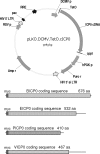
FIG. 1.
FIG. 1.
A map of the lentiviral vector used for expression of ICP0 and its related proteins and a summary of the coding sequences utilized. The upper part shows lentivirus plasmid vector pLKO.DCMV.TetO.cICP0 that includes the ICP0 cDNA. The key features of the lentivirus vector are noted: pac, HIV packaging sequence; RRE, Rev response element; RSV p, Rous sarcoma virus promoter; Amp r, ampicillin resistance; Puro-r, puromycin resistance; hGFPp, human phosphoglycerol kinase gene promoter; cppt, central polypyrimidine tract; DCMVp, truncated HCMV promoter/enhancer; TetO, tandem tetracycline operator sequences; LTR, long terminal repeat. The lower section shows the fragments inserted in place of the ICP0 cDNA in the above plasmid. Coding sequences are boxed, indicating the myc tag present in each and the length of the open reading frame; 3′ noncoding sequences are shown by lines. aa, amino acids.
FIG. 2.
FIG. 2.
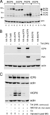
Expression of ICP0 family member proteins in inducible cell lines. (A) Cell lines transduced with lentivirus including the indicated open reading frames were treated with tetracycline, and then whole-cell extracts were prepared at 2, 4, and 6 h after induction, as shown. Expressed proteins were detected by Western blotting with an anti-myc tag antibody. (B) Comparative levels of ICP0 and BICP0 expression. Whole-cell extracts of uninduced and induced cells expressing untagged ICP0 (HA-cICP0) and myc-tagged ICP0 and BICP0 were analyzed by Western blotting for ICP0 (upper panel) and the myc tag (middle panel). The lower panel shows the actin loading control. (C) Instability of VICP0. HA-cICP0 and HA-VICP0 cells were left uninduced or were treated with tetracycline for 24 h. Samples were treated with MG132 (5 μM) for 2 h, and then replicate samples were maintained in tetracycline medium with cycloheximide (Cx) but without MG132 for 1, 2, and 4 h (all as indicted below the panels).
FIG. 3.
FIG. 3.
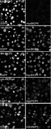
Expression of ICP0 family members in induced cell lines. Untreated HA-BICP0 cells (top row) and HA-BICP0, HA-EICP0, HA-PICP0, and HA-VICP0 cells treated with tetracycline for 4 h were stained for expression of the myc-tagged proteins. Images were captured on identical settings for the uninduced BICP0 cells and the induced BICP0, EICP0, and PICP0 samples. An enhanced setting was used for VICP0 cells because of the weaker signal (right-hand panel). The left-hand panels show the EGFP staining from expression of the EGFP-linked tetracycline repressor protein in all cells.
FIG. 4.
FIG. 4.
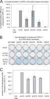
(A) Complementation of plaque formation efficiency of ICP0 null mutant HSV-1 by ICP0 family members. Cells expressing the indicated proteins were treated with tetracycline for 24 h and then used to titrate a stock of ICP0 null mutant HSV-1. The increase in titer in the ICP0-expressing cells in these experiments was an average of 330-fold greater than in the control HA-TetR cells. The data present average increases in titer over background in the other cell lines. The data show the mean of duplicate experiments, and the error bars show the range of the individual values. (B) Derepression of repressed HSV-1 genomes by induction of ICP0 family members and analysis of HA-TetR control cells and cells expressing the indicated proteins. Cells were infected (rows 2 and 3) or not (mock) with virus in1374 at an MOI of 5 PFU per cell at NPT, and then 24 h later the cells in row 3 were treated with tetracycline (0.1 μg/ml). The cells were stained for β-galactosidase activity the following day to detect reactivated transcription from the lacZ marker gene in the in1374 genome. Cells positive for reactivated transcription turn blue while negative cells give a clear background. (C) Quantification of the extent of derepression. Four random fields of view of the wells in row 3 of panel B were photographed, and then positive and negative cells were counted (approximately 250 cells per view). The proportions of positive (blue) cells in the induced wells are plotted with respect to the numbers in the HA-cICP0 samples. The bars show mean results from the different fields of view counted, with standard deviations. The background level of blue cells in HA-TetR cells representing those in which expression of the marker gene was not initially repressed is 0.5%. This constitutes the limit of detection, meaning that the assay extends over a 200-fold range.
FIG. 5.
FIG. 5.
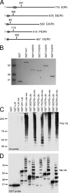
The RING domains of the ICP0 family members exhibit E3 ubiquitin ligase activity in vitro. (A) A representation of the ICP0 family member amino acid sequences, with their RING fingers marked by boxes. The arrows indicate the C-terminal limits of the sequences fused to GST and expressed in bacteria. (B) An image of a Coomassie-stained 4 to 12% Bis-Tris Novex gel loaded with 200 ng of each purified GST fusion protein used in the analysis. The details of the amino acid coordinates of the RING domains used and their purification are given in Materials and Methods. (C and D) Western blots showing the E3 ubiquitin (Ub) ligase activities of the ICP0 family proteins. Each purified GST fusion protein was incubated with either ubiquitin or a ubiquitin mix containing ubiquitin, E1 ubiquitin-activating enzyme, and E2 ubiquitin-conjugating enzyme UbcH5a, as indicated. For the blot analysis in panel C, samples containing wt ubiquitin and high-molecular-weight unlinked polyubiquitin chains were detected with P4D1 anti-ubiquitin antibody. The blot in panel D shows similar reactions conducted with methylated ubiquitin (Me Ub), which reveals multiple monoubiquitin-conjugated species of the GST fusion proteins produced by auto-ubiquitination.
FIG. 6.
FIG. 6.
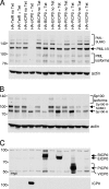
Degradation of PML and Sp100 induced by ICP0 family proteins. Control HA-TetR cells and cells transduced with lentiviruses encoding the ICP0 family members were treated with tetracycline for 24 h and analyzed by Western blotting for PML (A), Sp100 (B), and myc-tagged proteins or ICP0 (C). Uninduced samples (no Tet) of each cell line are shown for comparison. The panels show analysis of the same samples on replicate gels.
FIG. 7.
FIG. 7.
Analysis of the effects of ICP0 family members on ND10 proteins PML (two left columns), Sp100,(two middle columns) and hDaxx (two right columns) by immunofluorescence. The top row shows uninduced HA-BICP0 cells as controls. These are identical to uninduced samples from the other cell lines and HA-TetR parental cells (not shown). Rows 2 to 5 show typical images of groups of cells induced to express BICP0, EICP0, PICP0, and VICP0, as indicated, at 24 h after induction.
FIG. 8.
FIG. 8.
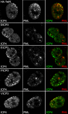
Inhibition of recruitment of PML to the sites of viral genomes by ICP0-related proteins during the early stages of HSV-1 infection. Control HA-TetR cells and cells induced by a 24-h tetracycline treatment to express BICP0 and the other family members were infected at a low MOI with ICP0 null mutant HSV-1. At 24 h after infection, the cells were fixed and stained for PML (red) and ICP4 (green). Representative images of cells close to the edge of developing plaques were selected to show the sites of parental viral genomes and early replication compartments (detected by concentrations of ICP4 in distinct foci). The recruitment of PML to these foci, obvious in the HA-TetR and VICP0-expressing cells (top and bottom rows), is inhibited by expression of BICP0, EICP0, and PICP0 (rows 2 to 4, as indicated).
FIG. 9.
FIG. 9.
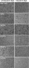
Viability of cells expressing ICP0 and related proteins. Control HA-TetR and cells expressing ICP0 and its related proteins were maintained in the absence (uninduced) or continuous presence (induced) of tetracycline for 6 days. The cells were then stained lightly with Giemsa and photographed.
FIG. 10.
FIG. 10.
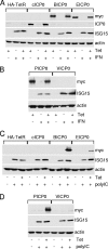
ICP0 family members do not impede induction of ISG15 expression by either IFN or poly(I:C) treatment. (A and B) The indicated cells lines were seeded in triplicate wells, two of which were treated with tetracycline for 24 h. The uninduced well and one of the induced wells were then treated with IFN-β for a further 24 h in the continuous presence of tetracycline. Whole-cell extracts were analyzed by Western blotting for ICP0, myc-tagged ICP0 family members, ISG15, and actin. (C and D) A similar experiment was performed except that poly(I:C) was used instead of interferon. In this experiment the negative-control lane for ISG15 induction was not treated with tetracycline and the control for ICP0 expression is not shown.

Similar articles

Cited by

References

    1. Barlow, P. N., B. Luisi, A. Milner, M. Elliott, and R. Everett. 1994. Structure of the C3HC4 domain by 1H-nuclear magnetic resonance spectroscopy. A new structural class of zinc-finger. J. Mol. Biol. 237:201-211. - PubMed
    1. Boldogkoi, Z., A. Braun, and I. Fodor. 2000. Replication and virulence of early protein 0 and long latency transcript deficient mutants of the Aujeszky's disease (pseudorabies) virus. Microbes Infect. 2:1321-1328. - PubMed
    1. Boutell, C., S. Sadis, and R. D. Everett. 2002. Herpes simplex virus type 1 immediate-early protein ICP0 and its isolated RING finger domain act as ubiquitin E3 ligases in vitro. J. Virol. 76:841-850. - PMC - PubMed
    1. Bowles, D. E., V. R. Holden, Y. Zhao, and D. J. O'Callaghan. 1997. The ICP0 protein of equine herpesvirus 1 is an early protein that independently transactivates expression of all classes of viral promoters. J. Virol. 71:4904-4914. - PMC - PubMed
    1. Bowles, D. E., S. K. Kim, and D. J. O'Callaghan. 2000. Characterization of the trans-activation properties of equine herpesvirus 1 EICP0 protein. J. Virol. 74:1200-1208. - PMC - PubMed

Publication types

MeSH terms

LinkOut - more resources