. 2010 Apr;84(7):3287-302.
doi: 10.1128/JVI.02020-09. Epub 2010 Jan 27.

Pharmacological manipulation of the akt signaling pathway regulates myxoma virus replication and tropism in human cancer cells

Affiliations

Pharmacological manipulation of the akt signaling pathway regulates myxoma virus replication and tropism in human cancer cells

Steven J Werden et al. J Virol. 2010 Apr.

Abstract

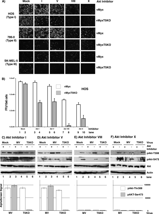
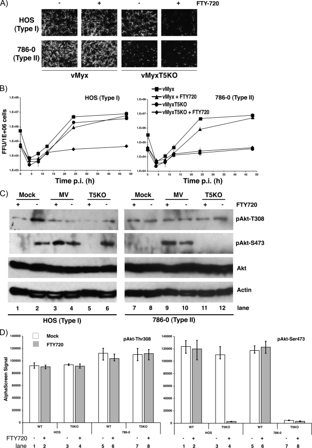
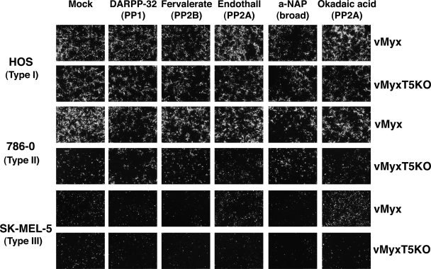
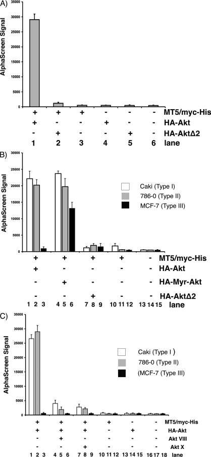
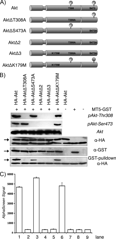
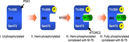
Viruses have evolved an assortment of mechanisms for regulating the Akt signaling pathway to establish a cellular environment more favorable for viral replication. Myxoma virus (MYXV) is a rabbit-specific poxvirus that encodes many immunomodulatory factors, including an ankyrin repeat-containing host range protein termed M-T5 that functions to regulate tropism of MYXV for rabbit lymphocytes and certain human cancer cells. MYXV permissiveness in these human cancer cells is dependent upon the direct interaction between M-T5 and Akt, which has been shown to induce the kinase activity of Akt. In this study, an array of compounds that selectively manipulate Akt signaling was screened and we show that only a subset of Akt inhibitors significantly decreased the ability of MYXV to replicate in previously permissive human cancer cells. Furthermore, reduced viral replication efficiency was correlated with lower levels of phosphorylated Akt. In contrast, the PP2A-specific phosphatase inhibitor okadaic acid promoted increased Akt kinase activation and rescued MYXV replication in human cancer cells that did not previously support viral replication. Finally, phosphorylation of Akt at residue Thr308 was shown to dictate the physical interaction between Akt and M-T5, which then leads to phosphorylation of Ser473 and permits productive MYXV replication in these human cancer cells. The results of this study further characterize the mechanism by which M-T5 exploits the Akt signaling cascade and affirms this interaction as a major tropism determinant that regulates the replication efficiency of MYXV in human cancer cells.


Figures

FIG. 1.
FIG. 1.
Effects of Akt inhibitors on MYXV replication in human cancer cells. (A) HOS, 786-0, and SK-MEL-5 cells were infected with either vMyx-gfp or vMyxT5KO-gfp in the absence (mock) or presence of various Akt inhibitors, and viral foci were detected by florescence microscopy at 48 hpi. Inhibitor concentrations are as stated in Materials and Methods. (B) Permissive HOS cells were infected with either vMyx-gfp (white bars) or vMyxT5KO-gfp (gray bars) at an MOI of 1 in the in the absence (mock) or presence of various Akt inhibitors, virus was collected at 48 hpi, and virus titer was determined using BGMK cells. Titers are expressed as numbers of FFU/106 cells and represent the means ± standard deviations of results from triplicate wells. (C to F) Representative Western blots (upper panels) and results of AlphaScreen SureFire assays (lower panels) illustrate Akt phosphorylation in cell lysates collected from HOS cells mock infected (lanes 1 and 2) or infected with either vMyx (MV; lanes 3 and 4) or vMyxT5KO (lanes 5 and 6) at MOI 3 in the presence (+) or absence (−) of various Akt inhibitors. Kinase activation of Akt is demonstrated by detection of specific phosphorylated forms pAkt-Thr308 (white bars) and pAkt-Ser473 (gray bars). Equal sample loading was confirmed by detection of total Akt and the housekeeping protein actin. For AlphaScreen SureFire, each sample was assayed in triplicate, and standard deviations are represented by the error bars.
FIG. 2.
FIG. 2.
Inhibition of vMyxT5KO replication by FTY720. (A) HOS, 786-0, and SK-MEL-5 human cancer cells were infected with either vMyx-gfp or vMyxT5KO-gfp in the absence (−) or presence (+) of FTY720, and at 48 hpi, viral focus formation was determined by florescence microscopy. (B) HOS (left) and 786-0 (right) cells were infected with either vMyx-gfp or vMyxT5KO-gfp at an MOI of 1 in the in the absence or presence of FTY720, and the titer of virus collected at various hpi was determined using BGMK cells. Titers are expressed as numbers of FFU/106 cells and represent the means ± standard deviations of results from triplicate wells. (C) Representative Western blots showing detection of Akt in cell lysates from HOS (left) and 786-0 (right) cells at 24 hpi following mock infection (lanes 1 and 2), or infection with either vMyx (lanes 3 and 4) or vMyxT5KO (lanes 5 and 6) at an MOI 3 in the in the presence (+) or absence (−) of FTY720. Kinase activation of Akt is demonstrated by detection of specific phosphorylated forms pAkt-Thr308 and pAkt-Ser473. Equal sample loading was confirmed by detection of actin. (D) Cell lysates of HOS cells prepared from those shown in panel C were assayed for Akt phospho-Thr308 (left) and Akt phospho-Ser473 (right) by using the standard AlphaScreen SureFire protocol. Each sample was assayed in triplicate, and standard deviations are represented by the error bars. Mock-treated cells are represented by white bars, whereas cells treated with FTY720 are represented by gray bars. WT, wild type.
FIG. 3.
FIG. 3.
PP2A-specific protein phosphatases rescue MYXV replication in nonpermissive type III cancer cells. MYXV focus formation was detected at 48 hpi by florescence microscopy in HOS, 786-0, and SK-MEL-5 cells infected with either vMyx-gfp or vMyxT5KO-gfp in the absence (mock) or presence of various protein phosphatase inhibitors. a-NAP, α-naphthyl acid phosphate, monosodium salt.
FIG. 4.
FIG. 4.
Combination of okadaic acid plus rapamycin enhances vMyxT5KO replication in type III cancer cells. The effect of okadaic acid on MYXV replication in HOS (lanes 1 to 4), 786-0 (lanes 5 to 8), and SK-MEL-5 (lanes 9 to 12) cells. All cells were either mock-treated (white bars) or preincubated with okadaic acid (gray bars) for 4 h prior to infection with vMyx-gfp (WT) or vMyxT5KO-gfp (T5KO), and at 48 hpi, focus formation was determined by florescence microscopy. (B) Single-step growth analysis of vMyx-gfp (open bars) and vMyxT5KO-gfp (gray bars) at 48 hpi in type III SK-MEL-5 cells. Prior to viral infection, cells were mock treated (lanes 1 and 2) or treated with either okadaic acid (lanes 3 and 4), rapamycin (lanes 5 and 6), or both okadaic acid and rapamycin (lanes 7 and 8). Titers are expressed as numbers of FFU/106 cells and represent the means ± standard deviations of results from triplicate wells. (C) Cell lysates were prepared from type III SK-MEL-5 cells mock infected (lanes 1 to 4) or infected with either vMyx (lanes 4 to 8) or vMyxT5KO (lanes 9 to 12) at an MOI of 3 in the absence (−) or presence (+) of okadaic acid and/or rapamycin.
FIG. 5.
FIG. 5.
The cellular environment regulates the interaction between Akt and M-T5. Cells were transiently transfected with the specified (+) combination of plasmids (MT5/myc-His, HA-Akt, HA-AktΔ2, and HA-Myr-Akt) in HEK293 (A) or Caki, 786-0, and MCF-7 (B and C) cell lines. Cell lysates were harvested after 48 h and preincubated with biotin conjugated anti-Myc antibody and donor/acceptor beads before an AlphaScreen assay was performed to determine binding. Each sample was assayed in triplicate, and the error bars represent standard deviations. (C) Cells were either mock treated or treated with Akt inhibitor VIII or X prior to collection of cell lysis and AlphaScreen analysis of M-T5 binding to Akt.
FIG. 6.
FIG. 6.
Phospho status of Akt residue Thr308 mediates M-T5 binding. (A) Schematic representation of the Akt constructs used during this study. Amino acid substitutions are indicated. All Akt constructs contain an amino-terminal HA tag. (B) The plasmid MT5-GST and the various HA-Akt constructs were coexpressed by in vitro-coupled transcription-translation and subjected to a GST pulldown assay. Precipitates and total lysates were resolved by SDS-PAGE and probed with anti-HA (α-HA) antibody to detect coprecipitated viral proteins (lower panels). Expression and phosphorylation of Akt proteins were confirmed by immunoblotting with phospho- and non-phospho-specific Akt antibodies and antibody against HA epitope (upper three panels). Furthermore, M-T5 expression was detected by an anti-GST-specific antibody. Bands of interest are represented by arrows. (C) Cell lysates were collected from transiently transfected HEK293 cells and assayed for protein-protein binding by AlphaScreen as described in the legend to Fig. 5.
FIG. 7.
FIG. 7.
Binding of M-T5 to cellular Akt induces phosphorylation of Akt and is a key restriction determinant for MYXV permissiveness in type II human cancer cells. Phosphorylation of Akt at residue Thr308 by PDK1 induces a conformational change to Akt, which allows M-T5 to bind. Binding of M-T5 to the hemi-phosphorylated Akt promotes the subsequent phosphorylation of Ser473 by mTORC2. Once fully phosphorylated, Akt activates various downstream signaling cascades important for MYXV replication in the human cancer cells. Type I cells are constitutively in conformation IV and are permissive for both wild-type MYXV and vMyxT5KO. Type III cells are constitutively trapped in conformation I and are nonpermissive for both wild-type MYXV and vMyxT5KO.

Similar articles

Cited by

References

    1. Ahn, J. Y., R. Rong, T. G. Kroll, E. G. Van Meir, S. H. Snyder, and K. Ye. 2004. PIKE (phosphatidylinositol 3-kinase enhancer)-A GTPase stimulates Akt activity and mediates cellular invasion. J. Biol. Chem. 279:16441-16451. - PubMed
    1. Alessi, D. R., M. Andjelkovic, B. Caudwell, P. Cron, N. Morrice, P. Cohen, and B. A. Hemmings. 1996. Mechanism of activation of protein kinase B by insulin and IGF-1. EMBO J. 15:6541-6551. - PMC - PubMed
    1. Azuma, H., S. Takahara, S. Horie, S. Muto, Y. Otsuki, and Y. Katsuoka. 2003. Induction of apoptosis in human bladder cancer cells in vitro and in vivo caused by FTY720 treatment. J. Urol. 169:2372-2377. - PubMed
    1. Azuma, H., S. Takahara, N. Ichimaru, J. D. Wang, Y. Itoh, Y. Otsuki, J. Morimoto, R. Fukui, M. Hoshiga, T. Ishihara, N. Nonomura, S. Suzuki, A. Okuyama, and Y. Katsuoka. 2002. Marked prevention of tumor growth and metastasis by a novel immunosuppressive agent, FTY720, in mouse breast cancer models. Cancer Res. 62:1410-1419. - PubMed
    1. Barnett, S. F., D. Defeo-Jones, S. Fu, P. J. Hancock, K. M. Haskell, R. E. Jones, J. A. Kahana, A. M. Kral, K. Leander, L. L. Lee, J. Malinowski, E. M. McAvoy, D. D. Nahas, R. G. Robinson, and H. E. Huber. 2005. Identification and characterization of pleckstrin-homology-domain-dependent and isoenzyme-specific Akt inhibitors. Biochem. J. 385:399-408. - PMC - PubMed

Publication types

MeSH terms

LinkOut - more resources