. 2010 Mar;16(3):598-609.
doi: 10.1261/rna.1852310. Epub 2010 Jan 27.

Idiosyncratically tuned switching behavior of riboswitch aptamer domains revealed by comparative small-angle X-ray scattering analysis

Affiliations

Idiosyncratically tuned switching behavior of riboswitch aptamer domains revealed by comparative small-angle X-ray scattering analysis

Nathan J Baird et al. RNA. 2010 Mar.

Erratum in

  • RNA. 2010 Jul;16(7):1447

Abstract

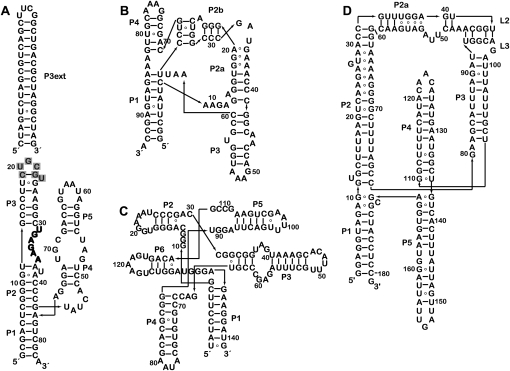
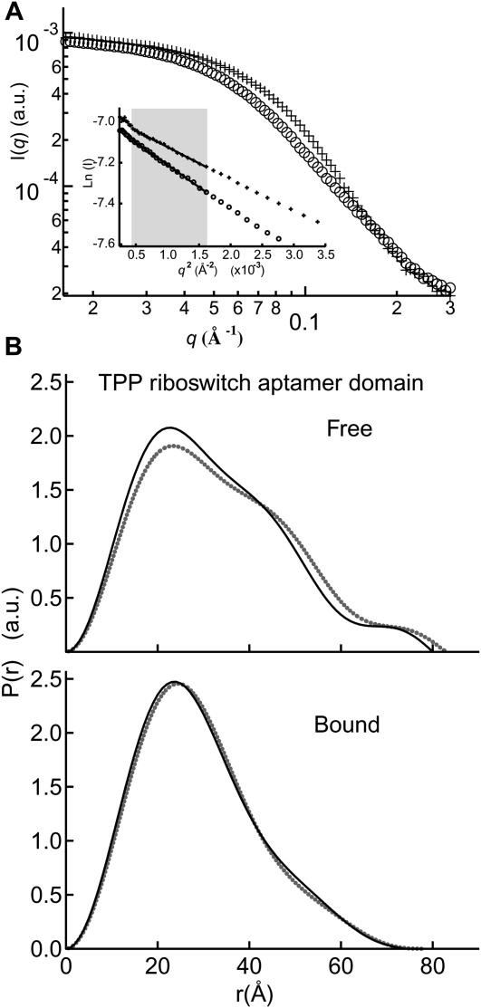
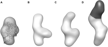
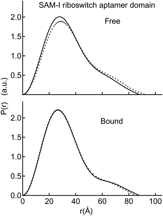
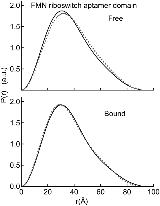
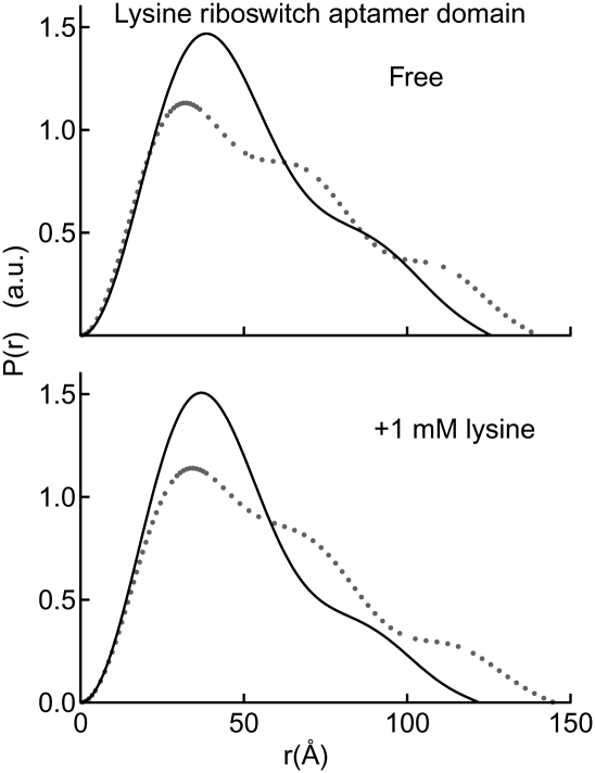
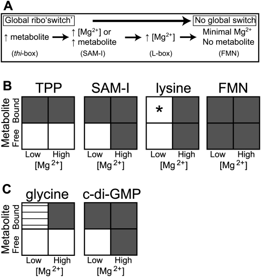
Riboswitches are structured mRNA elements that regulate gene expression upon binding specific cellular metabolites. It is thought that the highly conserved metabolite-binding domains of riboswitches undergo conformational change upon binding their cognate ligands. To investigate the generality of such a mechanism, we employed small-angle X-ray scattering (SAXS). We probed the nature of the global metabolite-induced response of the metabolite-binding domains of four different riboswitches that bind, respectively, thiamine pyrophosphate (TPP), flavin mononucleotide (FMN), lysine, and S-adenosyl methionine (SAM). We find that each RNA is unique in its global structural response to metabolite. Whereas some RNAs exhibit distinct free and bound conformations, others are globally insensitive to the presence of metabolite. Thus, a global conformational change of the metabolite-binding domain is not a requirement for riboswitch function. It is possible that the range of behaviors observed by SAXS, rather than being a biophysical idiosyncrasy, reflects adaptation of riboswitches to the regulatory requirements of their individual genomic context.


Figures

FIGURE 1.
FIGURE 1.
Sequence and schematic secondary structures of the riboswitch aptamer domains employed in this study. Secondary structure diagrams are organized similar to available 3D structures for riboswitch aptamers that bind (A) TPP, (B) SAM, (C) FMN, and (D) lysine. (P3ext) The helical extension added to P3 of the TPP aptamer domain (A). (Gray) The nucleotides removed from the wild-type sequence upon introduction of the extension. The T-loop between P2 and P3, which is responsible for recognition of the pyrimidine moiety of TPP, is highlighted in bold letters.
FIGURE 2.
FIGURE 2.
Metabolite-induced conformational switch in the TPP aptamer domain. (A) Sample raw scattering data and (insert) Guinier plots for determination of Rg; (circles) TPP-free; (crosses) TPP-bound. Most Guinier analyses in the present study were performed within the q range indicated by the shaded gray box. (B) P(r) plot detailing the differences in shape and maximum dimension between the free and bound conformations at 2.5 mM Mg2+ (gray dots) and 10 mM Mg2+ (black lines). (a.u.) Arbitrary units.
FIGURE 3.
FIGURE 3.
Low-resolution SAXS models of the TPP aptamer domain. (A) DAMMIN reconstruction of the bound conformation. The crystal structure is docked within it to demonstrate the agreement in size and shape. The correlation coefficient between the reconstruction and the envelope calculated from the crystal structure (Edwards and Ferré-D'Amaré 2006) is 0.89 (Wriggers et al. 1999). (B) Low-resolution model of the “free” form in the presence of 2.5 mM Mg2+. (C) Reconstruction of the free conformation in 1.5 mM Mg2+ is in general agreement with that at 2.5 mM Mg2+, although slightly expanded. (D) Low-resolution model of the P3ext variant (1.5 mM Mg2+) demonstrates an additional density (black) in the P3 region.
FIGURE 4.
FIGURE 4.
Conformational switching induced by Mg2+ and metabolite in the SAM-I aptamer domain. Two P(r) plots reveal changes in Dmax and global conformations between the free and bound states in the presence of 2.5 mM Mg2+ (gray dots) and 10 mM Mg2+ (black lines). The bound conformation is identical for the 2.5 mM and 10 mM Mg2+, rendering the 2.5 mM trace (gray dots) largely obstructed from view.
FIGURE 5.
FIGURE 5.
Global compaction of the FMN aptamer domain requires minimal Mg2+ and no metabolite. P(r) plots demonstrate the similarity of global conformation for the FMN aptamer domain in all conditions tested; free and bound in 1.5 mM (gray dots) and 10 mM Mg2+ (black lines).
FIGURE 6.
FIGURE 6.
Mg2+-dependent response of the lysine riboswitch aptamer domain. A P(r) plot clearly indicates compaction upon addition of highly stabilizing Mg2+ concentrations (10 mM; black lines) as compared to physiologic Mg2+ concentrations (1 mM Mg2+; gray dots). At 10 mM Mg2+, the global conformational change is induced only by Mg2+ (relative to the 1 mM Mg2+ condition) with virtually no difference between the free and bound states. At the low Mg2+ condition, the ligand is sub-saturating (see the text for details).
FIGURE 7.
FIGURE 7.
Alternative simplified schematics of global conformational responses induced by Mg2+ and metabolite. Each riboswitch investigated in this study exhibits idiosyncratic responses to the conditions probed. (A) Conditions are described that yield a globally compacted conformation for each aptamer domain as measured by Rg. (B) A matrix describing the degree of compaction relative to the low [Mg2+], no metabolite condition is diagrammed as follows: (white) expanded; (gray) fully compacted; (horizontal lines) partially compacted. (*) As indicated in the Discussion, lysine is present at sub-saturating concentration in our low-Mg2+ concentration condition. (C) The same matrix is presented for the glycine and cyclic diguanylate aptamer domain conformations, data adapted from Lipfert et al. (2007) and Kulshina et al. (2009), respectively. The low [Mg2+] reference point is chosen as 2.5 mM Mg2+ for TPP, SAM-I, lysine, and cyclic diguanylate aptamer domains; and 1.5 mM Mg2+ for the FMN and glycine aptamer domains. The high [Mg2+] reference point is 10 mM Mg2+ in all cases. Global changes are reported for conformations that exhibit differences in Rg > 1.5 Å.

Similar articles

Cited by

References

    1. Baird NJ, Westhof E, Qin H, Pan T, Sosnick TR. Structure of a folding intermediate reveals the interplay between core and peripheral elements in RNA folding. J Mol Biol. 2005;352:712–722. - PubMed
    1. Batey RT, Gilbert SD, Montange RK. Structure of a natural guanine-responsive riboswitch complexed with the metabolite hypoxanthine. Nature. 2004;432:411–415. - PubMed
    1. Chauhan S, Caliskan G, Briber RM, Perez-Salas U, Rangan P, Thirumalai D, Woodson SA. RNA tertiary interactions mediate native collapse of a bacterial group I ribozyme. J Mol Biol. 2005;353:1199–1209. - PubMed
    1. Dann CE, III, Wakeman CA, Sieling CL, Baker SC, Irnov I, Winkler WC. Structure and mechanism of a metal-sensing regulatory RNA. Cell. 2007;130:878–892. - PubMed
    1. Edwards TE, Ferré-D'Amaré AR. Crystal structures of the thi-box riboswitch bound to thiamine pyrophosphate analogs reveal adaptive RNA-small molecule recognition. Structure. 2006;14:1459–1468. - PubMed

Publication types

LinkOut - more resources