. 2010 Apr 9;285(15):11078-86.
doi: 10.1074/jbc.M109.034868. Epub 2010 Jan 27.

cAMP-stimulated protein phosphatase 2A activity associated with muscle A kinase-anchoring protein (mAKAP) signaling complexes inhibits the phosphorylation and activity of the cAMP-specific phosphodiesterase PDE4D3

Affiliations

cAMP-stimulated protein phosphatase 2A activity associated with muscle A kinase-anchoring protein (mAKAP) signaling complexes inhibits the phosphorylation and activity of the cAMP-specific phosphodiesterase PDE4D3

Kimberly L Dodge-Kafka et al. J Biol Chem. .

Abstract

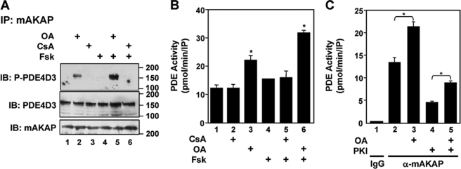
The concentration of the second messenger cAMP is tightly controlled in cells by the activity of phosphodiesterases. We have previously described how the protein kinase A-anchoring protein mAKAP serves as a scaffold for the cAMP-dependent protein kinase PKA and the cAMP-specific phosphodiesterase PDE4D3 in cardiac myocytes. PKA and PDE4D3 constitute a negative feedback loop whereby PKA-catalyzed phosphorylation and activation of PDE4D3 attenuate local cAMP levels. We now show that protein phosphatase 2A (PP2A) associated with mAKAP complexes is responsible for reversing the activation of PDE4D3 by catalyzing the dephosphorylation of PDE4D3 serine residue 54. Mapping studies reveal that a C-terminal mAKAP domain (residues 2085-2319) binds PP2A. Binding to mAKAP is required for PP2A function, such that deletion of the C-terminal domain enhances both base-line and forskolin-stimulated PDE4D3 activity. Interestingly, PP2A holoenzyme associated with mAKAP complexes in the heart contains the PP2A targeting subunit B56delta. Like PDE4D3, B56delta is a PKA substrate, and PKA phosphorylation of mAKAP-bound B56delta enhances phosphatase activity 2-fold in the complex. Accordingly, expression of a B56delta mutant that cannot be phosphorylated by PKA results in increased PDE4D3 phosphorylation. Taken together, our findings demonstrate that PP2A associated with mAKAP complexes promotes PDE4D3 dephosphorylation, serving both to inhibit PDE4D3 in unstimulated cells and also to mediate a cAMP-induced positive feedback loop following adenylyl cyclase activation and B56delta phosphorylation. In general, PKA.PP2A.mAKAP complexes exemplify how protein kinases and phosphatases may participate in molecular signaling complexes to dynamically regulate localized intracellular signaling.


Figures

FIGURE 1.
FIGURE 1.
An OA-sensitive phosphatase regulates mAKAP-associated PDE4D3. A, transfected HEK293 cells expressing both mAKAP and PDE4D3 were treated with either 300 μm OA or 500 μm cyclosporin A (CsA) for 30 min before stimulation with 5 μm Fsk for 10 min. The phosphorylation state of PDE4D3 present in mAKAP antibody immunoprecipitates (IP) was determined using a antibody specific for phosphorylated PDE4D3 Ser-54 (top panel). Total PDE4D3 (middle panel) and mAKAP (bottom panel) present in mAKAP antibody immunoprecipitates were detected using non-phospho-specific antibodies. Note that in these experiments mAKAP was GFP-tagged, and PDE4D3 was VSV and GFP-tagged, resulting in increased molecular weights. n = 3. IB, immunoblotted. B, PDE activity associated with mAKAP antibody immunoprecipitates prepared as in A was assayed using [3H]cAMP substrate. *, p < 0.05 compared with untreated cells (bar 1). C, endogenous protein complexes were isolated using control (IgG) or mAKAP-specific antibodies from clarified adult rat heart extracts (500 μg of total protein). PDE activity associated with the immunoprecipitates was assayed in the presence of 10 nm OA or 50 nm PKI. n = 3; *, p < 0.05.
FIGURE 2.
FIGURE 2.
PP2A is associated with the mAKAP scaffold in adult rat heart. A, phosphatase activity associated with protein complexes immunoprecipitated using mAKAP antibody from clarified adult rat heart extracts (500 μg of total protein) was assayed using 32P-labeled histone substrate in the absence or presence of 30 nm PP2A inhibitor 1 (30) and 100 nm PKA-phosphorylated PP1 inhibitor 1 (31). n = 3; *, p < 0.05. B and C, protein complexes were isolated from adult rat heart extracts (2 mg of total protein) using control (IgG) or mAKAP-specific antibody. 80 μg of total control extracts and 25% of the total immunoprecipitates were loaded onto the gel. PP2A (B) and PP1 (C) catalytic subunits were detected by immunoblotting (IB). n = 3.
FIGURE 3.
FIGURE 3.
PP2A binds a C-terminal mAKAP domain. A, schematic of mAKAP domains and GFP- and myc-tagged mAKAP proteins used in this paper. mAKAP fragments containing rat and human protein are drawn in black and gray, respectively. Hatched bars indicate the three spectrin repeat domains responsible for nuclear envelope targeting in myocytes (26). Binding sites are indicated for proteins known to bind mAKAP directly, including nesprin-1α (1074–1187) (11), ryanodine receptor (RyR2, 1217–1242) (39), PP2B (1286–1345) (14), PDE4D3 (1285–1833) (10), and PKA (2055–2072) (26). For reference, the binding site for 3-phosphoinositide-dependent kinase-1 (PDK1, mAKAP residues 227–232) (28) is indicated, although this protein is not discussed further in this paper. The stippled bar marks the PP2A binding site. The first and last residues of each fragment are indicated. B, purified GST-PP2A-A subunit fusion protein was incubated with extracts prepared from HEK293 cells expressing the indicated GFP-mAKAP fusion protein and pulled down using glutathione resin. GFP-mAKAP fragments were detected in the pulldowns (25% loaded, top panel) and the extracts (5% loaded, bottom panel) using a GFP antibody. n = 3. IB, immunoblotting. C, myc-tagged mAKAP fragments were expressed in HEK293 cells, and phosphatase binding was detected by immunoprecipitation using control (IgG) or myc-tag antibody followed by phosphatase assay using 32P-labeled histone substrate. n = 3. *, p < 0.05 compared with the other samples.
FIGURE 4.
FIGURE 4.
PP2A association with mAKAP·PDE4D3 complexes is required for inhibition of PDE4D3 phosphorylation. A, HEK293 cells expressing (VSV- and GFP-tagged) PDE4D3 and myc-tagged mAKAP 1286–2312 or 1286–2083 lacking the PP2A binding site were treated with 300 μm OA for 30 min before stimulation with 5 μm Fsk for 10 min. Protein complexes were immunoprecipitated (IP) using myc-tagged antibody in the presence of phosphatase inhibitors. The phosphorylation state of co-immunoprecipitated PDE4D3 was determined using an antibody specific for phosphorylated PDE4D3 Ser-54 (P-PDE4D3, top panel). Total PDE4D3, myc-mAKAP, and PP2A-C subunit present in the immunoprecipitates were detected using non-phospho-specific antibodies (bottom three panels). n = 3. IB, immunoblotting. B, PDE activity associated with myc-antibody immunoprecipitates isolated from additional cells treated as in A was assayed using [3H]cAMP. n = 3. *, p < 0.05 compared with bar 1.
FIGURE 5.
FIGURE 5.
mAKAP-bound PP2A contains B56δ subunit and is cAMP-activated. A, protein complexes were immunoprecipitated from clarified adult rat heart extracts (500 μg of total protein) using control (IgG) or mAKAP-specific antibody as in Fig. 2B and assayed for associated phosphatase activity. As indicated, the immunoprecipitates were preincubated with no addition or with 50 μm CPT-cAMP, 10 nm OA, or 50 nm PKI for 5 min before the addition of [32P]histone substrate. n = 3; *, p < 0.05. B, endogenous protein complexes were immunoprecipitated from adult heart extract (2 mg of total protein) with B56δ and control (IgG) antibodies in 80 μg of extract, and 25% of the immunoprecipitates were loaded onto the gel. mAKAP was detected by immunoblotting (IB). n = 3. C, FLAG-tagged B56δ and/or GFP-tagged mAKAP were expressed in HEK293 cells. Protein complexes were immunoprecipitated (IP) using a mAKAP antibody. B56δ in the immunoprecipitates (25% loaded) and total extracts (5% loaded) was detected by immunoblotting with a FLAG antibody. n = 3. D, phosphatase activity associated with mAKAP-antibody immunoprecipitates prepared as in C was assayed using 32P-labeled histone substrate. n = 3. E, HEK293 cells expressing mAKAP and B56δ were treated with 5 μm Fsk and 10 μm IBMX (Fsk/IBMX) for 10 min before immunoprecipitation of protein complexes with mAKAP antibody. Phosphatase activity associated with the immunoprecipitates was assayed using [32P]histone substrate. n = 3. Note that PP2A B56δ and C subunit binding to mAKAP was not affected by Fsk/IBMX (see Fig. 6).
FIGURE 6.
FIGURE 6.
Phosphorylation of B56δ by PKA increases mAKAP-associated PP2A activity. A, B56δ is phosphorylated on serine residues 53, 68, 81, and 566 by PKA (25). B56δ wild-type or alanine-substituted at all four PKA sites (S4A) was co-expressed in HEK293 cells with wild-type (WT) mAKAP or a full-length mAKAP mutant lacking the PKA binding site (ΔPKA; cf. Fig. 3A). After stimulation with 5 μm Fsk and 50 μm IBMX, protein complexes were immunoprecipitated (IP) with mAKAP antibody, and associated proteins were detected by immunoblotting (IB) with B56δ, mAKAP, and PP2A-C antibodies (bottom three panels). PKA phosphorylation of B56δ was detected by immunoblotting with a B56δ phospho-Ser-566-specific antibody (P-B56δ, top panel). n = 3. B, immunoprecipitates prepared as in A were assayed for associated phosphatase activity. n = 3; * p < 0.05.
FIGURE 7.
FIGURE 7.
Phosphorylation of B56δ by PKA enhances the dephosphorylation of mAKAP-associated PDE3D3. A, HEK293 cells expressing (GFP-tagged) mAKAP, (VSV- and GFP-tagged) PDE4D3 and either wild-type B56δ or B56δ S4A mutant at the PKA phosphorylation sites were treated as indicated with 300 μm OA for 30 min before stimulation for 10 min with 5 μm Fsk. Protein complexes were immunoprecipitated (IP) with mAKAP antibody in the presence of phosphatase inhibitors. The phosphorylation state of PDE4D3 present in the immunoprecipitates was determined using an antibody specific for phosphorylated PDE4D3 Ser-54 (top panel). Total PDE4D3, mAKAP, B56δ, and PP2A-C protein present in the immunoprecipitates were detected using non-phospho-specific antibodies (lower four panels). n = 3. IB, immunoblotting. B, PDE activity associated with protein complexes isolated from additional cells treated as in A was assayed using [3H]cAMP. n = 3; *, p < 0.05 compared with bar 1.
FIGURE 8.
FIGURE 8.
PKA and PP2A associated with mAKAP complexes coordinately regulate PDE4D3 activity and cAMP degradation. PKA is composed of two regulatory and two catalytic subunits. mAKAP-bound PP2A contains A, B56δ, and C (catalytic) subunits. A, in unstimulated cells, basal PP2A activity maintains PDE4D3 dephosphorylation, presumably allowing for a more rapid rise in cAMP levels in response to subsequent agonist than if PDE4D3 were phosphorylated and activated. At the same time, basal PDE4D3 activity should maintain low local levels of cAMP, preventing spurious signaling. B, Gs-coupled receptor stimulation induces cAMP synthesis, exceeding the rate of cAMP degradation by PDE4D3 and activating mAKAP-bound PKA. PKA phosphorylates and activates both PDE4D3 and PP2A. PDE4D3 activation should limit peak cAMP levels as well as accelerate the rate of cAMP clearance after GPCR down-regulation. In contrast, PP2A activation opposes PDE4D3 phosphorylation by PKA, attenuating cAMP degradation and contributing to greater, longer lasting cAMP signals.

Similar articles

Cited by

References

    1. Hayes J. S., Brunton L. L., Mayer S. E. (1980) J. Biol. Chem. 255, 5113–5119 - PubMed
    1. Brunton L. L., Hayes J. S., Mayer S. E. (1979) Nature 280, 78–80 - PubMed
    1. Keely S. L. (1977) Res. Commun. Chem. Pathol. Pharmacol. 18, 283–290 - PubMed
    1. Keely S. L. (1979) Mol. Pharmacol. 15, 235–245 - PubMed
    1. Steinberg S. F., Brunton L. L. (2001) Annu. Rev. Pharmacol. Toxicol. 41, 751–773 - PubMed

Publication types

LinkOut - more resources