

Cell surface of Lactococcus lactis is covered by a protective polysaccharide pellicle
- PMID: 20106971
- PMCID: PMC2856253
- DOI: 10.1074/jbc.M109.082958
Cell surface of Lactococcus lactis is covered by a protective polysaccharide pellicle
Abstract
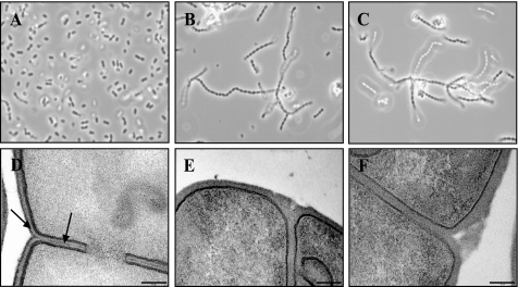
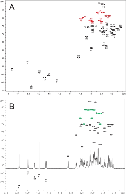
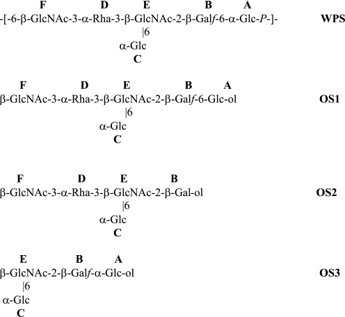
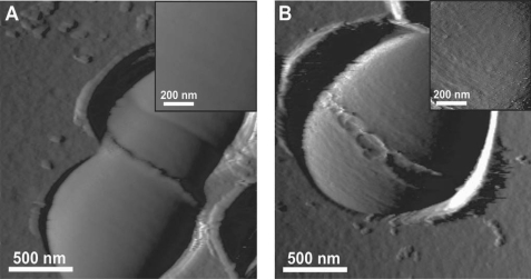
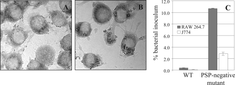
In Gram-positive bacteria, the functional role of surface polysaccharides (PS) that are not of capsular nature remains poorly understood. Here, we report the presence of a novel cell wall PS pellicle on the surface of Lactococcus lactis. Spontaneous PS-negative mutants were selected using semi-liquid growth conditions, and all mutations were mapped in a single chromosomal locus coding for PS biosynthesis. PS molecules were shown to be composed of hexasaccharide phosphate repeating units that are distinct from other bacterial PS. Using complementary atomic force and transmission electron microscopy techniques, we showed that the PS layer forms an outer pellicle surrounding the cell. Notably, we found that this cell wall layer confers a protective barrier against host phagocytosis by murine macrophages. Altogether, our results suggest that the PS pellicle could represent a new cell envelope structural component of Gram-positive bacteria.
Figures
Similar articles
-
Another Brick in the Wall: a Rhamnan Polysaccharide Trapped inside Peptidoglycan of Lactococcus lactis.mBio. 2017 Sep 12;8(5):e01303-17. doi: 10.1128/mBio.01303-17. mBio. 2017. PMID: 28900021 Free PMC article.
-
A dual-chain assembly pathway generates the high structural diversity of cell-wall polysaccharides in Lactococcus lactis.J Biol Chem. 2019 Nov 15;294(46):17612-17625. doi: 10.1074/jbc.RA119.009957. Epub 2019 Oct 3. J Biol Chem. 2019. PMID: 31582566 Free PMC article.
-
PBP2b Mutations Improve the Growth of Phage-Resistant Lactococcus cremoris Lacking Polysaccharide Pellicle.Appl Environ Microbiol. 2023 Jun 28;89(6):e0210322. doi: 10.1128/aem.02103-22. Epub 2023 May 24. Appl Environ Microbiol. 2023. PMID: 37222606 Free PMC article.
-
Bacillus anthracis cell envelope components.Curr Top Microbiol Immunol. 2002;271:87-113. doi: 10.1007/978-3-662-05767-4_5. Curr Top Microbiol Immunol. 2002. PMID: 12224525 Review.
-
Adhesin receptors of human oral bacteria and modeling of putative adhesin-binding domains.J Ind Microbiol. 1995 Sep;15(3):176-85. doi: 10.1007/BF01569823. J Ind Microbiol. 1995. PMID: 8519475 Review.
Cited by
-
Cell Wall Glycans Mediate Recognition of the Dairy Bacterium Streptococcus thermophilus by Bacteriophages.Appl Environ Microbiol. 2018 Nov 15;84(23):e01847-18. doi: 10.1128/AEM.01847-18. Print 2018 Dec 1. Appl Environ Microbiol. 2018. PMID: 30242010 Free PMC article.
-
Recognition of gram-positive intestinal bacteria by hybridoma- and colostrum-derived secretory immunoglobulin A is mediated by carbohydrates.J Biol Chem. 2011 May 13;286(19):17239-47. doi: 10.1074/jbc.M110.209015. Epub 2011 Mar 21. J Biol Chem. 2011. PMID: 21454510 Free PMC article.
-
Molecular mechanisms underlying the structural diversity of rhamnose-rich cell wall polysaccharides in lactococci.J Biol Chem. 2024 Jan;300(1):105578. doi: 10.1016/j.jbc.2023.105578. Epub 2023 Dec 16. J Biol Chem. 2024. PMID: 38110036 Free PMC article.
-
Genetics of Lactococci.Microbiol Spectr. 2019 Jul;7(4):10.1128/microbiolspec.gpp3-0035-2018. doi: 10.1128/microbiolspec.GPP3-0035-2018. Microbiol Spectr. 2019. PMID: 31298208 Free PMC article. Review.
-
PilVax - a novel peptide delivery platform for the development of mucosal vaccines.Sci Rep. 2018 Feb 7;8(1):2555. doi: 10.1038/s41598-018-20863-7. Sci Rep. 2018. PMID: 29416095 Free PMC article.
References
Publication types
MeSH terms
Substances
LinkOut - more resources
Full Text Sources
Other Literature Sources
Molecular Biology Databases
