. 2010 Mar 26;285(13):9962-9970.
doi: 10.1074/jbc.M109.087098. Epub 2010 Jan 27.

Crystal structure of Get4-Get5 complex and its interactions with Sgt2, Get3, and Ydj1

Affiliations

Crystal structure of Get4-Get5 complex and its interactions with Sgt2, Get3, and Ydj1

Yi-Wei Chang et al. J Biol Chem. .

Abstract

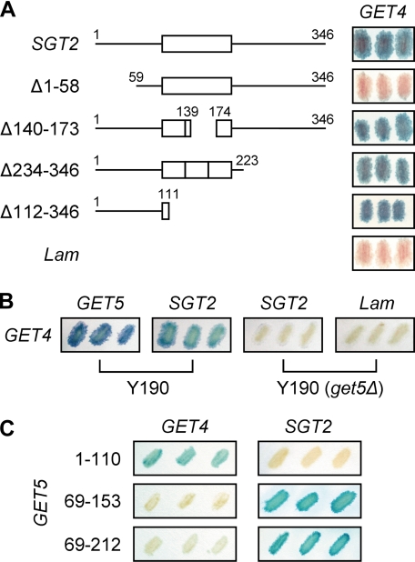
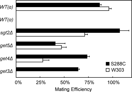
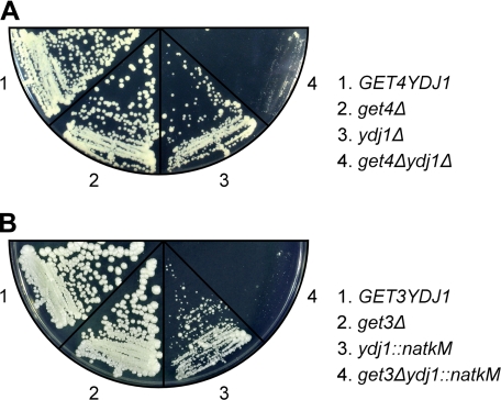
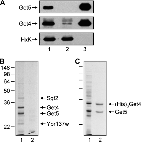
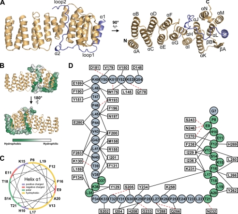
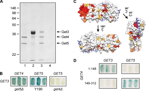
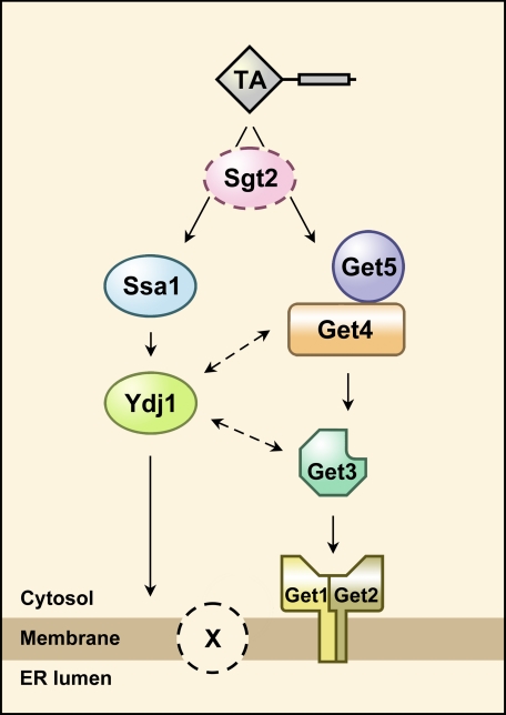
Get3, Get4, and Get5 in Saccharomyces cerevisiae participate in the insertion of tail-anchored proteins into the endoplasmic reticulum membrane. We elucidated the interaction between Get4 and Get5 and investigated their interaction with Get3 and a tetratricopeptide repeat-containing protein, Sgt2. Based on co-immunoprecipitation and crystallographic studies, Get4 and Get5 formed a tight complex, suggesting that they constitute subunits of a larger complex. In contrast, although Get3 interacted physically with the Get4-Get5 complex, low amounts of Get3 co-precipitated with Get5, implying a transient interaction between Get3 and Get4-Get5. Sgt2 also interacted with Get5, although the amount of Sgt2 that co-precipitated with Get5 varied. Moreover, GET3, GET4, and GET5 interacted genetically with molecular chaperone YDJ1, suggesting that chaperones might also be involved in the insertion of tail-anchored proteins.


Figures

FIGURE 1.
FIGURE 1.
Yeast two-hybrid assays to assess the interaction between different pairs of proteins among Sgt2, Get4, and Get5. A, interaction of SGT2 and its fragments (in pAS2-1 vector) with GET4 (in pACT2 vector) was determined, and the blue filters are shown. Human lamin C-(66–230) in pAS2-1 (Lam) was used as a control. B, interaction of GET4 with SGT2 was determined using yeast strain Y190 and Y190 with GET5 deleted; the blue filters are shown. C, interaction of GET4 and SGT2 with fragments of GET5 (in either pAS2-1 or pACT2) was determined, and the blue filters are shown. The ubiquitin-like domain of Get5 ranges from residues 74–173.
FIGURE 2.
FIGURE 2.
Effect of deletions of GET or SGT2 genes on mating efficiency. The mating efficiency of the mutants having the W303 or S288C genetic background was quantified. Data represent the means ± S.E. of three independent determinations. WT, wild type.
FIGURE 3.
FIGURE 3.
Genetic interaction of YDJ1 with GET4 and GET5. Diploid yeast strains heterozygous for YDJ1 and GET4 (A) or for YDJ1 and GET3 (B) were sporulated, and tetrads were dissected. Growth fitness assays were performed on tetrads with all four possible combinations. Here, an equal number of cells was streaked onto YPD plates and allowed to grow at 30 °C for 3–4 days before being photographed.
FIGURE 4.
FIGURE 4.
Get4 and Get5 form a complex. A, yeast lysates were prepared and utilized for immunoprecipitation with anti-Get5. The immunoprecipitates were subjected to gel electrophoresis and then blotted with anti-Get5, anti-Get4, and anti-hexokinase (HxK). Lanes 1, 2, and 3 represent the lysate diluted 1:2000, flow-through diluted 1:2000, and protein bound to antibodies diluted 1:200, respectively. B, the protein immunoprecipitated with anti-Get5 was subjected to gel electrophoresis, and the gel was stained with Coomassie Brilliant Blue. Lanes 1 and 2 represent immunoprecipitates from wild-type and get5Δ lysates, respectively. C, Get5 co-purifies with His-tagged Get4 using Ni2+-NTA resin. Lanes 1 and 2 represent the soluble fraction of the bacterial lysates and the purified protein, respectively.
FIGURE 5.
FIGURE 5.
Crystal structure of Get4-Get5N. A, ribbon diagrams for the Get4-Get5N complex structure. The structure of Get4 in the complex (gold) and Get5N (blue) is shown. The left and right panels are the front orthogonal view and top orthogonal view, respectively. B, hydrophobicity of the Get5N surface. The surface hydrophobicity of Get5N is shown by a color gradient from green (hydrophobic) to white (hydrophilic). Get4 is shown as the gold ribbons. C, helical plot of the α1 in Get5N. The basic, acidic, polar, and hydrophobic residues are indicated in blue, red, green, and yellow, respectively. D, interactions between Get5N and Get4. Residues of Get5N and Get4 are displayed by circles and rectangles, respectively. The α-helices and loops of Get5N are colored in green and blue, respectively. Residues participating in intermolecular interactions are represented by black solid lines (hydrophobic interactions) or red dotted lines (hydrogen bonds).
FIGURE 6.
FIGURE 6.
Interaction of the Get4-Get5 complex with Get3. A, recombinant Get4-Get5 complex (lane 1) was incubated with purified Get3 (lane 2), and the mixtures were immunoprecipitated with anti-Get5. Get3 co-precipitated in the presence of the Get4-Get5 complex (lane 3) but not in its absence (lane 4). B, yeast two-hybrid interactions of GET3 with GET4 in Y190(get5Δ) and of GET3 with GET5 in Y190 and Y190(get4Δ) are shown. C, conserved residues on the Get4 surface. Residue conservation in Get4 determined by sequence alignment (supplemental Fig. S5) is shown by a surface diagram with a color gradient from yellow (70% conserved) to red (100% identical). Get5N (blue) is shown as ribbons with stick side chains. D, the yeast two-hybrid assays were used to dissect the interaction of Get4 with Get3 and Get5. The N-terminal region (residues 1–148) and C-terminal region (residues 149–312) of Get4 are responsible for interacting with Get3 and Get5, respectively.
FIGURE 7.
FIGURE 7.
Model for the insertion of tail-anchored proteins. Besides the known pathway involving GET proteins, there is a chaperone-dependent pathway for the insertion of tail-anchored proteins. However, the role of Sgt2, if any, in this process needs to be elucidated. It also remains to be determined whether there are additional membrane proteins (indicated by X) participating in the insertion of tail-anchored proteins.

References

    1. Becker J., Walter W., Yan W., Craig E. A. (1996) Mol. Cell. Biol. 16, 4378–4386 - PMC - PubMed
    1. D'Andrea L. D., Regan L. (2003) Trends Biochem. Sci. 28, 655–662 - PubMed
    1. Wegele H., Haslbeck M., Reinstein J., Buchner J. (2003) J. Biol. Chem. 278, 25970–25976 - PubMed
    1. Hainzl O., Wegele H., Richter K., Buchner J. (2004) J. Biol. Chem. 279, 23267–23273 - PubMed
    1. Young J. C., Hoogenraad N. J., Hartl F. U. (2003) Cell 112, 41–50 - PubMed

Publication types

MeSH terms

Associated data