Review
. 2010 Apr;298(4):R851-61.
doi: 10.1152/ajpregu.00002.2010. Epub 2010 Jan 27.

Mechanisms of proximal tubule sodium transport regulation that link extracellular fluid volume and blood pressure

Affiliations
Review

Mechanisms of proximal tubule sodium transport regulation that link extracellular fluid volume and blood pressure

Alicia A McDonough. Am J Physiol Regul Integr Comp Physiol. 2010 Apr.

Erratum in

  • Am J Physiol Regul Integr Comp Physiol. 2010 May;298(5):R1448

Abstract

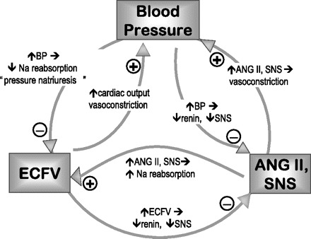
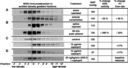
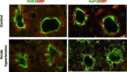
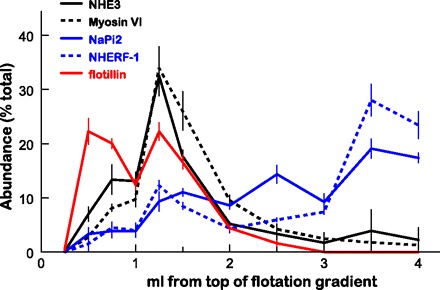
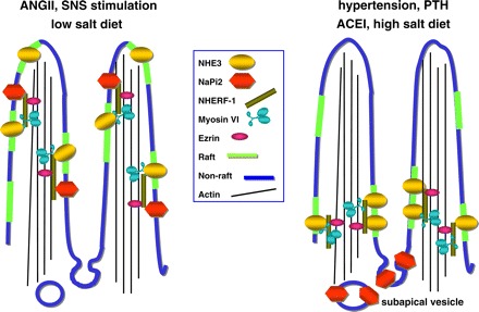
One-hundred years ago, Starling articulated the interdependence of renal control of circulating blood volume and effective cardiac performance. During the past 25 years, the molecular mechanisms responsible for the interdependence of blood pressure (BP), extracellular fluid volume (ECFV), the renin-angiotensin system (RAS), and sympathetic nervous system (SNS) have begun to be revealed. These variables all converge on regulation of renal proximal tubule (PT) sodium transport. The PT reabsorbs two-thirds of the filtered Na(+) and volume at baseline. This fraction is decreased when BP or perfusion pressure is increased, during a high-salt diet (elevated ECFV), and during inhibition of the production of ANG II; conversely, this fraction is increased by ANG II, SNS activation, and a low-salt diet. These variables all regulate the distribution of the Na(+)/H(+) exchanger isoform 3 (NHE3) and the Na(+)-phosphate cotransporter (NaPi2), along the apical microvilli of the PT. Natriuretic stimuli provoke the dynamic redistribution of these transporters along with associated regulators, molecular motors, and cytoskeleton-associated proteins to the base of the microvilli. The lipid raft-associated NHE3 remains at the base, and the nonraft-associated NaPi2 is endocytosed, culminating in decreased Na(+) transport and increased PT flow rate. Antinatriuretic stimuli return the same transporters and regulators to the body of the microvilli associated with an increase in transport activity and decrease in PT flow rate. In summary, ECFV and BP homeostasis are, at least in part, maintained by continuous and acute redistribution of transporter complexes up and down the PT microvilli, which affect regulation of PT sodium reabsorption in response to fluctuations in ECFV, BP, SNS, and RAS.


Figures

Fig. 1.
Fig. 1.
Feedback relationship between blood pressure (BP), ANG II, and sympathetic nervous system (SNS), and extracellular fluid volume (ECFV).
Fig. 2.
Fig. 2.
Distinct patterns of sodium hydrogen exchanger (NHE3) trafficking in response to different stimuli. Density distribution pattern of NHE3 before and after a treatment was assessed by fractionating renal cortex on sorbitol density gradients. A constant volume of each of the 12 fractions collected was analyzed by immunoblot with anti-NHE3. A: when mean arterial pressure (MAP) is elevated by arterial constriction, NHE3 moves out of low-density microvilli-enriched membranes to higher-density membranes found at the base of the microvilli. This redistribution is associated with a decrease in proximal tubule (PT) Na+ reabsorption (48) and a decrease in Na+/H+-exchanger activity (57), and an increase in end PT flow rate (10). B: when MAP is elevated by acute phenol injury, which stimulates sympathetic outflow to the kidneys, NHE3 redistributes from higher-density membranes found at the base of the microvilli into the low-density microvilli membranes. This redistribution is associated with an increase in NHE activity and decrease in PT flow rate (23). C: when the angiotensin-converting enzyme inhibitor (ACEI) captopril is infused at a dose that does not change MAP, renal blood flow (RBF), or glomerular filtration rate (GFR), NHE3 moves out of low-density microvilli-enriched membranes to higher-density membranes found at the base of the microvilli. This retraction is associated with an increase in PT flow rate (21). D: when ANG II is added to the captopril infusate at a dose that does not change MAP, RBF, or GFR, NHE3 distribution and PT flow rate return to the baseline distribution (34). (Note: minor variations in the absolute density distributions from one study to another are secondary to person-to-person variation in making and collecting gradients). A dashed line between fractions 5 and 6 is provided as a visual aid to facilitate detecting the redistribution patterns.
Fig. 3.
Fig. 3.
Differential redistribution of NHE3 and NaPi2 during acute hypertension. In this study (48), the endocytic compartment of the PT was labeled by intravenous injection of horseradish peroxidase (HRP), and rats were sham operated (control), or blood pressure was increased for 20 min (acute hypertension). Kidneys were fixed in situ, sectioned, and double labeled with either polyclonal anti-NHE3 or anti-NaPi2 (both green), and with monoclonal anti-HRP (red). Left: NHE3 is retracted from the body to the base of the microvilli during acute hypertension, with no evidence that NHE3 moves into endocytic tracer HRP-labeled compartment. Right: NaPi2 is retracted from the body of the microvilli and is subsequently colocalized with the endocytic tracer HRP (yellow indicates colocalization).
Fig. 4.
Fig. 4.
Flotation gradient profiles of NHE3, NaPi2, and associated proteins. Renal cortex total membranes treated with Triton X-100 were fractionated on OptiPrep gradients (35). The top 1.5 ml was collected as six 250-μl samples to delineate the lipid raft domain, and the remainder of the gradient was collected as five 500-μl aliquots. A constant volume of each fraction from untreated Sprague-Dawley rats was assayed by immunoblot. Profiles for NHE3 (solid black), myosin VI (dashed black), NaPi2 (solid blue) NHERF-1 (dashed blue), and the lipid raft marker flotillin (solid red) are compared. Data are expressed as the percentage of the total signal in all 11 fractions (means ± SE).
Fig. 5.
Fig. 5.
Schematic representing response of renal PT sodium transporters NaPi2 and Na+/H+ exchanger 3 (NHE3) to natriuretic and antinatriuretic stimuli. With the antinatriuretic stimuli, ANG II, SNS simulation/phenol injury and low-salt diet, both NaPi2 and NHE3 are located in the body of the microvilli: NHE3, DPPIV, and myosin VI are enriched in lipid rafts (green), while NaPi2 is enriched in nonlipid rafts (blue) along with NHE regulatory factor 1 (NHERF-1) and ezrin (35). Both NaPi2 and NHE3 have been reported to be associated with the PDZ adaptor protein NHERF-1 and are presumably tethered to the cytoskeleton via ezrin (42). How myosin VI is tethered to the transporters and the actin core remains to be determined. With natriuretic stimuli, such as elevated arterial or perfusion pressure, ACEI, high-salt diet or PTH, NHE3, and NaPi2 are translocated, within the plane of the membrane, out of the body of the microvilli. During PTH treatment, NHERF-1 is phosphorylated and dissociates from NaPi2 (41), presumably allowing the cotransporter to move into the intermicrovillar cleft, where it is endocytosed to subapical dense apical tubules and endosomes (48). In contrast, NHE3 remains in a domain at the base of the microvilli along with myosin VI, both localized to lipid rafts (48, 49).

Similar articles

Cited by

References

    1. Ahmed F, Kemp BA, Howell NL, Siragy HM, Carey RM. Extracellular renal guanosine cyclic 3'5'-monophosphate modulates nitric oxide and pressure-induced natriuresis. Hypertension 50: 958–963, 2007 - PubMed
    1. Ally S, Larson AG, Barlan K, Rice SE, Gelfand VI. Opposite-polarity motors activate one another to trigger cargo transport in live cells. J Cell Biol 187: 1071–1082, 2009 - PMC - PubMed
    1. Azuma KK, Balkovetz DF, Magyar CE, Lescale-Matys L, Zhang Y, Chambrey R, Warnock DG, McDonough AA. Renal Na+/H+ exchanger isoforms and their regulation by thyroid hormone. Am J Physiol Cell Physiol 270: C585–C592, 1996 - PubMed
    1. Bianchi G. Genetic variations of tubular sodium reabsorption leading to “primary” hypertension: from gene polymorphism to clinical symptoms. Am J Physiol Regul Integr Comp Physiol 289: R1536–R1549, 2005 - PubMed
    1. Biemesderfer D, Mentone SA, Mooseker M, Hasson T. Expression of myosin VI within the early endocytic pathway in adult and developing proximal tubules. Am J Physiol Renal Physiol 282: F785–F794, 2002 - PubMed

Publication types

MeSH terms

LinkOut - more resources