. 2010 Apr;17(4):660-7.
doi: 10.1128/CVI.00358-09. Epub 2010 Jan 27.

In situ immunolocalization and stage-dependent expression of a secretory serine protease in Leishmania donovani and its role as a vaccine candidate

Affiliations

In situ immunolocalization and stage-dependent expression of a secretory serine protease in Leishmania donovani and its role as a vaccine candidate

Rajdeep Choudhury et al. Clin Vaccine Immunol. 2010 Apr.

Abstract

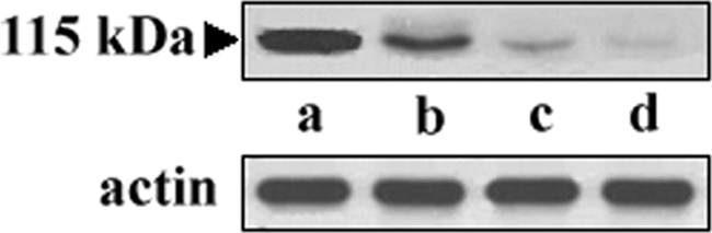
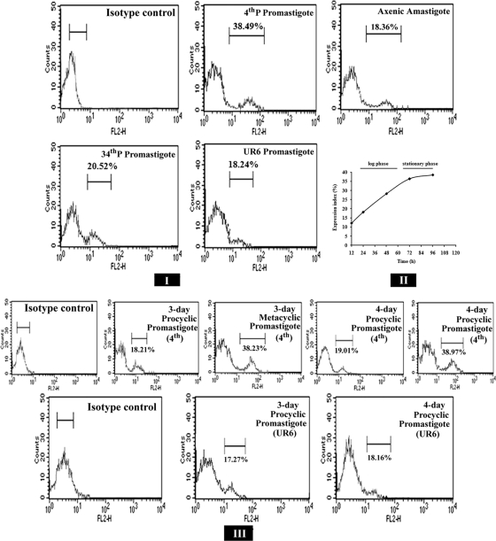
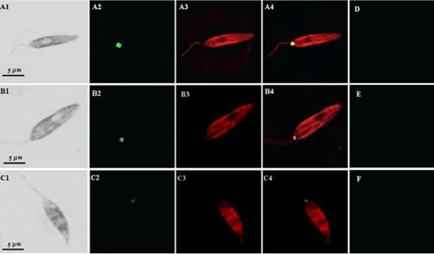
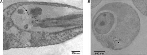
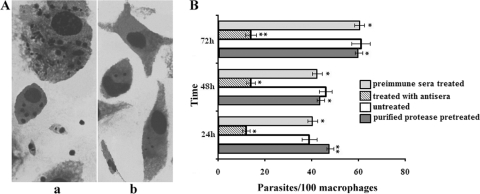
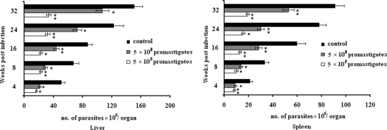
Proteases have been found to play essential roles in many biological processes, including the pathogenesis of leishmaniasis. Most parasites rely on their intracellular and extracellular protease repertoire to invade and multiply in mammalian host cells. However, few studies have addressed serine proteases in Leishmania and their role in host pathogenesis. Here we report the intracellular distribution of a novel L. donovani secretory serine protease in the flagellar pocket, as determined by immunogold labeling. Flow cytometry and confocal immunofluorescence analysis revealed that the expression of the protease diminishes sequentially from virulent to attenuated strains of this species and is also highly associated with the metacyclic stage of L. donovani promastigotes. The level of internalization of parasites treated with the anti-115-kDa antibody into host macrophages was significantly reduced from that of non-antibody-treated parasites, suggesting that this serine protease probably plays a role in the infection process. In vivo studies confirmed that this serine protease is a potential vaccine candidate. Altogether, the 115-kDa serine protease might play vital roles in L. donovani pathogenesis and hence could be recognized as a potential candidate for drug design.


Figures

FIG. 1.
FIG. 1.
Western blot analysis of the 115-kDa protease of L. donovani. Cell-free cultured supernatants from early-passage promastigotes and whole-cell extracts from virulent, avirulent, and attenuated strains of L. donovani were loaded directly onto an SDS gel and used for immunoblot detection with the anti-115-kDa serine protease antibody. Lane a, purified protease (2 μg protein) from the culture supernatant of L. donovani; lane b, whole-cell extract of early-passage (4th-P) L. donovani promastigotes (100 μg protein); lane c, whole-cell extract of late-passage (34th-P) L. donovani promastigotes (100 μg protein); lane d, whole-cell extract of UR6 promastigotes (100 μg protein). Actin was used as a loading control to normalize the data.
FIG. 2.
FIG. 2.
Flow cytometric analysis of expression of the 115-kDa extracellular serine protease. (I) Fluorescence histograms showing the expression levels of the 115-kDa protease in 4th-P, 34th-P, and UR6 promastigotes and axenic amastigotes of L. donovani. (II) Expression of the 115-kDa protease of 4th-P promastigotes of L. donovani at different phases of growth. The expression index measures the percentage of cells of the total population recognized by the anti-115-kDa antibody. (III) Fluorescence histograms showing the expression levels of the 115-kDa protease at procyclic and metacyclic stages of virulent promastigotes and at procyclic stages of attenuated UR6 promastigotes at 72 h and 96 h of culture.
FIG. 3.
FIG. 3.
Immunolocalization and differential expression of the 115-kDa serine protease in L. donovani. Early-passage (A1 to A4), late-passage (B1 to B4), and UR6 (C1 to C4) promastigotes of L. donovani were fixed and stained with an anti-115-kDa antibody and an anti-gp63 antibody as described in Materials and Methods. The immunofluorescence images of the promastigotes labeled for the 115-kDa serine protease are shown in the green channel (A2, B2, and C2), and those labeled for gp63 are shown in the red channel (A3, B3, and C3). Merged images (yellow channel) are shown in panels A4, B4, and C4. The corresponding phase-contrast images are shown on the left (A1, B1, and C1). No fluorescence was detected in similar preparations reacted with the preimmune serum (D, E, and F).
FIG. 4.
FIG. 4.
Intracellular localization of the 115-kDa serine protease of L. donovani by immunogold electron microscopy. Promastigotes (A) and amastigotes (B) of L. donovani were labeled with an anti-115-kDa serine protease antibody prior to detection with a gold-conjugated secondary antibody. Electron microscopy images show the presence of gold particles in the flagellar pocket regions of the parasites (arrows).
FIG. 5.
FIG. 5.
Effect of an antibody against the 115-kDa serine protease on host cell invasion by early-passage L. donovani promastigotes. (A) Microscopic images of macrophages incubated with non-antibody-treated (a) or antibody-treated (b) parasites. (B) Parasites were treated with the antibody for 1 h, after which unbound antibodies were washed out, and parasites were incubated with macrophages for 24 h, 48 h, or 72 h. In another set of experiments, macrophages were pretreated with the purified protease and were then used in the macrophage invasion assay. The internalized parasites were counted after staining with Giemsa stain. Results of corresponding control experiments in which parasites were not treated with the antibody or were treated with preimmune serum are also shown. Reversal of invasion inhibition was observed for macrophages pretreated with the purified 115-kDa serine protease. Results are means ± standard deviations for four independent experiments. Asterisks indicate significance levels (*, P < 0.05; **, P < 0.01).
FIG. 6.
FIG. 6.
Evaluation of protection against visceral leishmaniasis in BALB/c mice after immunization with the purified 115-kDa serine protease. At different time points postimmunization, parasite burdens in the livers and spleens of mice challenged with 5 × 10⁵ or 5 × 10⁸ virulent L. donovani promastigotes were quantified in Giemsa-stained preparations of impression smears. The results are expressed as the number of parasites per organ. Each experiment was performed at least three times, and the results are means ± standard deviations. Asterisks indicate significance levels (*, P < 0.05; **, P < 0.01).

Similar articles

Cited by

References

    1. Alexander, J., and D. G. Russel. 1992. The interaction of Leishmania species and macrophages. Adv. Parasitol. 31:175-254. - PubMed
    1. Atayde, V. D., M. Cortez, R. Souza, J. F. da Silveira, and N. Yoshida. 2007. Expression and cellular localization of molecules of the gp82 family in Trypanosoma cruzi metacyclic trypomastigotes. Infect. Immun. 75:3264-3270. - PMC - PubMed
    1. Bates, P. A. 1994. Complete developmental cycle of Leishmania mexicana in axenic culture. Parasitology 108:1-9. - PubMed
    1. Breton, C. B., T. Blisnick, H. Jouin, J. C. Barale, T. Rabilloud, G. Langsley, and L. H. P. da Silva. 1992. Plasmodium chabaudi p68 serine protease activity required for merozoite entry into mouse erythrocytes. Proc. Natl. Acad. Sci. U. S. A. 89:9647-9651. - PMC - PubMed
    1. Breton, M., M. J. Tremblay, M. Ouellette, and B. Papadopoulou. 2005. Live nonpathogenic parasitic vector as a candidate vaccine against visceral leishmaniasis. Infect. Immun. 73:6372-6382. - PMC - PubMed

Publication types

MeSH terms

LinkOut - more resources