. 2010 May;91(Pt 5):1311-4.
doi: 10.1099/vir.0.019307-0. Epub 2010 Jan 27.

The HR motif in the RNA-dependent RNA polymerase L protein of Chandipura virus is required for unconventional mRNA-capping activity

Affiliations

The HR motif in the RNA-dependent RNA polymerase L protein of Chandipura virus is required for unconventional mRNA-capping activity

Tomoaki Ogino et al. J Gen Virol. 2010 May.

Abstract

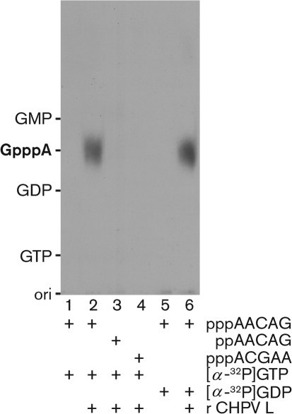
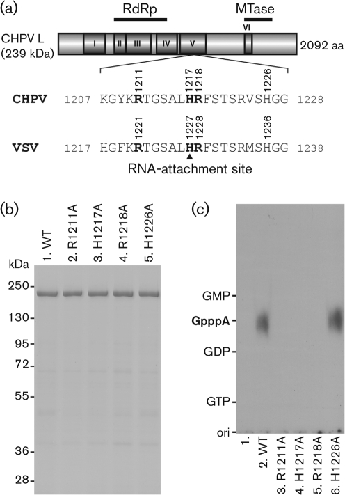
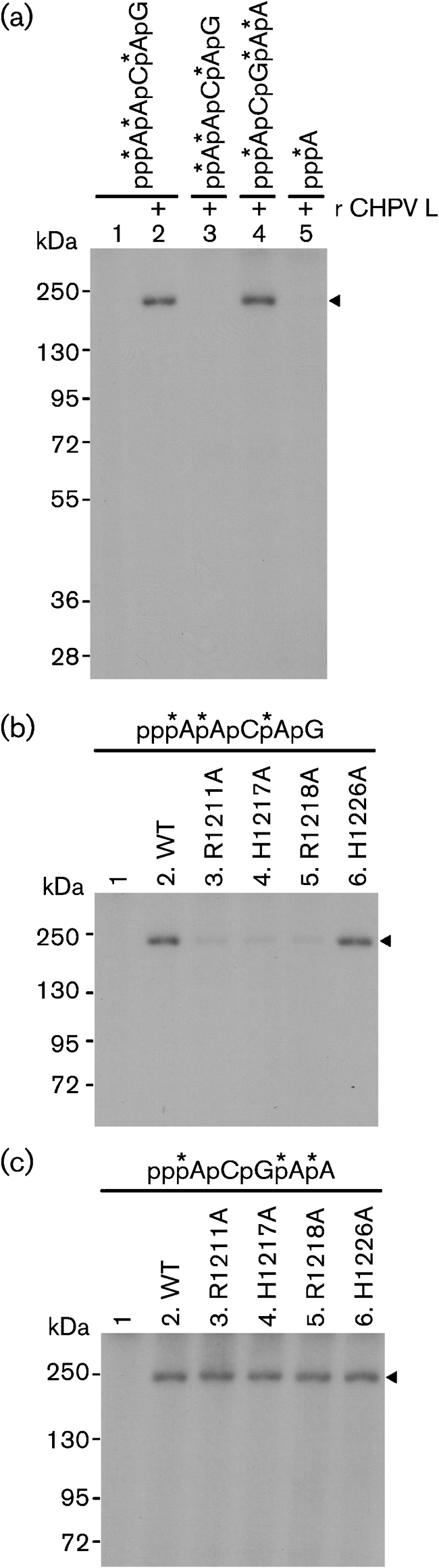
Chandipura virus (CHPV) is an emerging human pathogen associated with acute encephalitis and is related closely to vesicular stomatitis virus (VSV), a prototype rhabdovirus. Here, we demonstrate that the RNA polymerase L protein of CHPV exhibits a VSV-like RNA:GDP polyribonucleotidyltransferase (PRNTase) activity, which transfers the 5'-monophosphorylated (p-) viral mRNA start sequence to GDP to produce a capped RNA, and that the conserved HR motif in the CHPV L protein is essential for the PRNTase activity. Interestingly, the CHPV L protein was found to form two distinct SDS-resistant complexes with the CHPV mRNA and leader RNA start sequences; mutations in the HR motif significantly reduced the formation of the former complex (a putative covalent enzyme-pRNA intermediate in the PRNTase reaction), but not the latter complex. These results suggest that the rhabdoviral L proteins universally use the active-site HR motif for the PRNTase reaction at the step of the enzyme-pRNA intermediate formation.


Figures

Fig. 1.
Fig. 1.
RNA-capping activity of the CHPV L protein. The recombinant CHPV L protein (0.3 μg) was subjected to RNA-capping reactions with the indicated substrates. Calf intestine alkaline phosphatase- and nuclease P1-resistant products were analysed by PEI–cellulose thin-layer chromatography followed by autoradiography. Lanes 1 and 5 indicate no L protein. The positions of standard marker compounds, visualized under UV light at a wavelength of 254 nm, are shown on the left.
Fig. 2.
Fig. 2.
The HR motif is essential for the PRNTase activity of the CHPV L protein. (a) A schematic structure of the CHPV L protein (2092 aa) is shown, with six amino acid sequence blocks (I–VI) conserved in the NNS RNA viral L proteins. The positions of the putative RdRp and cap methyltransferase (MTase) domains are indicated. The local sequence containing the HR motif in the CHPV L protein is compared with that in the VSV L protein (H1227, the covalent RNA-attachment site). (b) Wild-type (WT) and mutant CHPV L proteins with indicated amino acid substitutions (0.7 μg) were analysed by SDS-PAGE (7.5 % gel) followed by staining with Coomassie brilliant blue. The positions of marker proteins are indicated on the left. (c) The WT and mutant CHPV L proteins (0.3 μg) were subjected to RNA-capping reactions with pppAACAG and [α-32P]GDP as substrates. Lane 1 indicates no L protein.
Fig. 3.
Fig. 3.
The HR motif in the CHPV L protein is required for the formation of the enzyme–pRNA intermediate in the PRNTase reaction. (a) Recombinant CHPV L protein (0.3 μg) was incubated with the indicated mono- or polyribonucleotide labelled with 32P (shown by asterisks), and then analysed by SDS-PAGE (7.5 % gel) followed by autoradiography. Note that all nucleotide probes had the same specific radioactivities. Lane 1 indicates no L protein. The position of covalent L–RNA complexes is indicated by the arrowhead. (b, c) The WT and mutant CHPV L proteins (0.3 μg) were incubated with pppAACAG [mRNA start sequence (b)] or pppACGAA [leader RNA start sequence (c)] labelled with 32P (shown by asterisks). The resulting covalent L–RNA complexes were analysed by SDS-PAGE (7.5 % gel) followed by autoradiography. Lane 1 indicates no L protein.

References

    1. Bourhy, H., Kissi, B. & Tordo, N. (1993). Molecular diversity of the Lyssavirus genus. Virology 194, 70–81. - PubMed
    1. Chadha, M. S., Arankalle, V. A., Jadi, R. S., Joshi, M. V., Thakare, J. P., Mahadev, P. V. & Mishra, A. C. (2005). An outbreak of Chandipura virus encephalitis in the eastern districts of Gujarat state, India. Am J Trop Med Hyg 73, 566–570. - PubMed
    1. Chang, S. H., Hefti, E., Obijeski, J. F. & Bishop, D. H. (1974). RNA transcription by the virion polymerases of five rhabdoviruses. J Virol 13, 652–661. - PMC - PubMed
    1. Furuichi, Y. & Shatkin, A. J. (2000). Viral and cellular mRNA capping: past and prospects. Adv Virus Res 55, 135–184. - PMC - PubMed
    1. Hoffmann, B., Schutze, H. & Mettenleiter, T. C. (2002). Determination of the complete genomic sequence and analysis of the gene products of the virus of spring viremia of carp, a fish rhabdovirus. Virus Res 84, 89–100. - PubMed

Publication types

LinkOut - more resources