. 2010 May;298(5):C1265-75.
doi: 10.1152/ajpcell.00341.2009. Epub 2010 Jan 27.

Collagen I matrix turnover is regulated by fibronectin polymerization

Affiliations

Collagen I matrix turnover is regulated by fibronectin polymerization

Feng Shi et al. Am J Physiol Cell Physiol. 2010 May.

Abstract

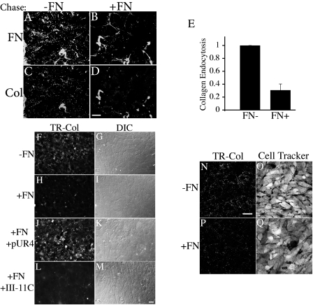
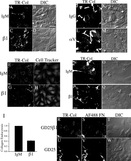
Extracellular matrix (ECM) remodeling occurs during normal homeostasis and also plays an important role during development, tissue repair, and in various disease processes. ECM remodeling involves changes in the synthesis, deposition, and degradation of ECM molecules. ECM molecules can be degraded extracellularly, as well as intracellularly following endocytosis. Our data show that the ECM protein fibronectin is an important regulator of ECM remodeling. We previously showed that agents that inhibit the polymerization of fibronectin into ECM fibrils promote the loss of preexisting fibronectin matrix and accelerate fibronectin endocytosis and degradation. In this paper we show that inhibition of fibronectin polymerization leads to the loss of collagen I matrix fibrils and a corresponding increase in the levels of endocytosed collagen I. In contrast, manipulations that stabilize fibronectin matrix fibrils, such as caveolin-1 depletion, stabilize collagen I matrix fibrils and cause a decrease in ECM collagen I endocytosis. Our data also show that endocytosis of ECM collagen I is regulated by both beta1 integrins and Endo180/urokinase plasminogen activator associated protein (uPARAP). Unexpectedly, Endo180/uPARAP was also shown to promote the endocytosis of fibronectin from the ECM. These data demonstrate that fibronectin polymerization regulates the remodeling of ECM collagen I, in part, by regulating collagen I endocytosis. Furthermore, these data show that processes that regulate ECM deposition coordinately regulate the removal of proteins from the ECM. These data highlight the complexity of ECM remodeling. This multifaceted regulatory process may be important to ensure tight regulation of ECM fibronectin and collagen I levels.


Figures

Fig. 1.
Fig. 1.
Endocytosis of collagen I and fibronectin (FN) from the extracellular matrix (ECM). AF: FN-null myofibroblasts (MFs) were incubated with Texas red (TR)-collagen I and AF488-FN overnight. Cells were then either fixed immediately (AC) or washed and then incubated for 24 h in media lacking fibronectin and collagen I (DF). Collagen I (A) and fibronectin (B) fibrils elaborated by cells after the overnight pulse were visualized with an Olympus fluorescence microscope. The same fields of view are shown in A and B. The corresponding differential interference contrast (DIC) picture is shown in C. Endocytosed TR-collagen I (D) and AF488-FN (E) were visualized after 24 h of chase. F is an overlay of D and E. Bar, 20 μm. G–I and K–M: FN-null MFs (GI) or SMC (KM) were seeded onto preassembled matrix containing TR-collagen I and AF488-FN and cultured for 24 h at 37°C. Endocytosed TR-collagen I (G, K) and AF488-FN (H, L) are shown. I, M are overlay images; the yellow staining indicates colocalized collagen and fibronectin. Scale bar, GI, 20 μm; KM, 10 μm. For DI and KM, a large portion of the extracellular FN and collagen fibrils were lost during the chase. Confocal images were taken in a plane of focus that would best show the intracellular vesicles. J: in vitro polymerized collagen gels (containing TR-collagen) were formed on top of the cell layer as described in materials and methods. After 48 h, the collagen gel was gently removed. The cells were then treated with 0.02% trypan blue for 5 min before fixation to quench extracellular fluorescence. Endocytosed TR-collagen is shown. Scale bar, 10 μm. DM are confocal images.
Fig. 2.
Fig. 2.
FN polymerization reduces collagen I endocytosis from matrix. AM: FN-null MFs were incubated with TR-collagen I and AF488-FN overnight. Cells were washed and then incubated (chased) for 24 h in media lacking labeled fibronectin and collagen (A, C, F) or containing 10 μg/ml unlabeled FN (B, D, H, J, L). During the chase, cells were also incubated in the absence (AI) or presence of 500 nM pUR4 (J, K) or III-11C (L, M) peptides. G, I, K, and M are DIC images corresponding to the left panel. For AD, a large portion of the extracellular FN and collagen fibrils were lost during the chase. Confocal images were taken in a plane of focus that would best show the intracellular vesicles. For F, H, J, and L, cells were treated with 0.02% trypan blue for 5 min before fixation to quench extracellular fluorescence. Scale bar, AD, 20 μm; FM, 60 μm. E: quantitation of matrix collagen I endocytosis. Fluorescent images were quantitated as described in materials and methods. The fluorescence intensity is reported as intensity per field of view. The graph in E shows the fold change relative to the mean fluorescence intensity of endocytosed TR-collagen I in cells chased in the absence of FN, which was set equal to 1 in each individual experiment. N–Q: confluent cultures of FN-null MFs were incubated in the absence (N, O) or presence (P, Q) of 10 μg/ml fibronectin for 12 h. The cell culture media was removed, and 2 mg/ml collagen (containing 20 μg/ml TR-collagen) was added on top of the cells and allowed to polymerize as described in materials and methods. Cells were cultured in the absence (N, O) or presence (P, Q) of 10 μg/ml fibronectin for an additional 48 h. The collagen gel was gently removed. The cells were then treated with 0.02% trypan blue for 5 min to quench extracellular fluorescence. Scale bar, 10 μm. AD and NQ are confocal images.
Fig. 3.
Fig. 3.
β1 Integrins regulate collagen I endocytosis from the ECM. A–M: FN-null MFs were seeded onto preassembled cell-derived matrices containing TR-collagen I in the presence of 25 μg/ml β1 integrin inhibitory (C, D, G, and H), 50 μg/ml αv inhibitory (L, M) or isotype control antibodies (A, B, E, F: IgM; J and K: IgG). Cells were cultured at 37°C for 22–36 h before fixation. A and C are confocal images taken in a plane of focus to best show extracellular collagen fibrils. E, G, J, and L are confocal images taken in a plane of focus to best visualize endocytosed collagen I; some extracellular fibrils can also be seen in these images. The cell outlines were visualized by loading cells with CellTracker green (F, H) or by DIC imaging (B, D, K, and M). Scale Bar, AD, 20 μm; EH, 20 μm; JM, 20 μm. I: quantitation of ECM collagen I endocytosis from preassembled matrix. Fluorescent images were quantitated as described in materials and methods. The fluorescence intensity is reported as intensity per unit area. The graph in I shows the fold change relative to the mean fluorescence intensity of endocytosed TR-collagen I in cells treated with isotype control IgM, which was set equal to 1 for each individual experiment. N–Q: smooth muscle cells (SMC) were seeded onto preassembled cell-derived matrices containing TR-collagen I in the presence of 25 μg/ml β1 integrin inhibitory (P, Q) or isotype control antibodies (N, O). Cells were incubated for 39 h before fixation. For N, P confocal images were taken in a plane of focus to best show intracellular vesicular fluorescence. The presence of the β1 integrin inhibitory antibody causes a reduction in intracellular vesicles and a preservation of matrix fibrils, as shown in P. Corresponding differential interference contrast (DIC) images are shown in O and Q. Scale Bar, 20 μm. R–W: GD25 β1 integrin null cells (UW) and GD25 β1 reexpressing cells (RT) were seeded onto preassembled cell-derived matrices containing TR-collagen I and AF488-FN and cultured for 24 h at 37°C. Confocal images were taken in a plane of focus to best show intracellular vesicular fluorescence. The absence of β1 integrins results in reduced intracellular vesicles and increased stability of extracellular matrix fibrils, as shown in U and V. TR-collagen (R, U) and AF488-FN (S, V) are shown. T and W are corresponding DIC images. Scale bar, 25 μm.
Fig. 4.
Fig. 4.
Expression of Endo180 in various cell lines. A: RT-PCR was performed as described in materials and methods. Endo180 mRNA was detected in FN-null MFs (lane 1), GD25 (lane 2), and GD25-β1 reexpressing cells (lane 3). B: cell lysates were prepared from Endo180 null cells (null), wild-type control cells (WT), GD25 (β1-), and GD25-β1 reexpressing (β1+) cells and analyzed by Western blot analysis using an antibody to Endo180. ERK was used as loading control. Molecular mass standards in kDa are shown on the left.
Fig. 5.
Fig. 5.
Endocytosis of ECM collagen I is reduced in caveolin-1 knockdown cells. FN-null MFs expressing caveolin-1 small interfering RNA (siRNA) (shcav, DF) or control cells (shluc, AC) were seeded onto preassembled matrix containing TR-collagen I and AF488-FN. Cells were incubated for 40 h at 37°C before fixation. Cells expressing caveolin-1 siRNA show enhanced stability of extracellular matrix collagen (E) and FN fibrils (D). Few intracellular vesicles were detected in caveolin-1 siRNA expressing cells (D, E). There was a paucity of ECM fibrils and a corresponding increase in intracellular TR-collagen I (B) and AF488-FN (A) in cells expressing control siRNA. Arrows in B point to internalized TR-collagen I. C and F are corresponding DIC images. A, B, D, and E are confocal images. Bar, 20 μm. G: quantitation of ECM collagen I endocytosis in caveolin-1 knockdown cells. Long-term pulse-chase assay were performed in FN-null MFs expressing caveolin-1 siRNA (shcav) or control cells (shluc) as described in materials and methods. Cells were chased for 14–18 h in the absence of exogenously added fibronectin and collagen I. Fluorescent images were quantitated as described in materials and methods. The fluorescence intensity is reported as intensity per field of view. The graph in G shows the fold change relative to the mean fluorescence intensity of endocytosed TR-collagen I in cells expressing control siRNA (shluc), which was set equal to 1 in each individual experiment.
Fig. 6.
Fig. 6.
Endo180 regulates the endocytosis of collagen I from the ECM. Endo180 null (DF) or WT control (AC) cells were seeded onto preassembled matrix containing TR-collagen I and AF488-FN. Cells were cultured for 24 h before fixation. TR-collagen I (B, E) and AF488-FN (A, D) are shown. Confocal images were taken in a plane of focus that would best show intracellular vesicles. Arrows in B point to abundant internalized TR-collagen I in WT control cells. Abundant internalized fibronectin is also seen in WT cells (A). There were few intracellular vesicles and a corresponding increase in the stability of fibrillar fibronectin and collagen I in Endo180 null cells (D,E). C and F are corresponding DIC images. Bar, 20 μM.
Fig. 7.
Fig. 7.
GM6001 inhibits collagen I matrix turnover and endocytosis. FN-null MFs were incubated with 10 μg/ml AF488-FN and 5 μg/ml (AI) or 10 μg/ml (JM) TR-collagen I overnight. A and B show extracellular matrix fibrils present in cells after the overnight incubation with fluorescently labeled fibronectin (A) or collagen (B) (“pulse”, AC). Cells were washed and then incubated for 24 h in media lacking fibronectin and collagen I but containing 20 μM GM6001, a metalloproteinase inhibitor (GI, L, M), or vehicle control (DF, J, K). Images in D, E, G, and H were taken in a plane of focus to best show extracellular matrix fibrils. In JM, cells were treated with 0.02% trypan blue to quench extracellular fluorescence prior to fixation as described in materials and methods. AF488-FN is shown in A, D, and G; TR-collagen I is shown in B, E, H, J, and L. Corresponding phase (C, F, I) or DIC (K, M) images are shown. AI: bar, 80 μm. JM: bar, 20 μm.

Similar articles

Cited by

References

    1. Ala-aho R, Kahari VM. Collagenases in cancer. Biochimie 87: 273–286, 2005 - PubMed
    1. Bae E, Sakai T, Mosher DF. Assembly of exogenous fibronectin by fibronectin-null cells is dependent on the adhesive substrate. J Biol Chem 279: 35749–35759, 2004 - PubMed
    1. Barilla ML, Carsons SE. Fibronectin fragments and their role in inflammatory arthritis. Semin Arthritis Rheum 29: 252–265, 2000 - PubMed
    1. Berk BC, Fujiwara K, Lehoux S. ECM remodeling in hypertensive heart disease. J Clin Invest 117: 568–575, 2007 - PMC - PubMed
    1. Bhide VM, Laschinger CA, Arora PD, Lee W, Hakkinen L, Larjava H, Sodek J, McCulloch CA. Collagen phagocytosis by fibroblasts is regulated by decorin. J Biol Chem 280: 23103–23113, 2005 - PubMed

Publication types

MeSH terms

LinkOut - more resources