. 2010 Jan 27;30(4):1238-49.
doi: 10.1523/JNEUROSCI.4464-09.2010.

Retinophilin is a light-regulated phosphoprotein required to suppress photoreceptor dark noise in Drosophila

Affiliations

Retinophilin is a light-regulated phosphoprotein required to suppress photoreceptor dark noise in Drosophila

Kirk L Mecklenburg et al. J Neurosci. .

Abstract

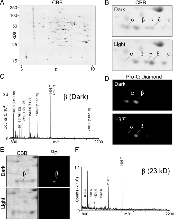
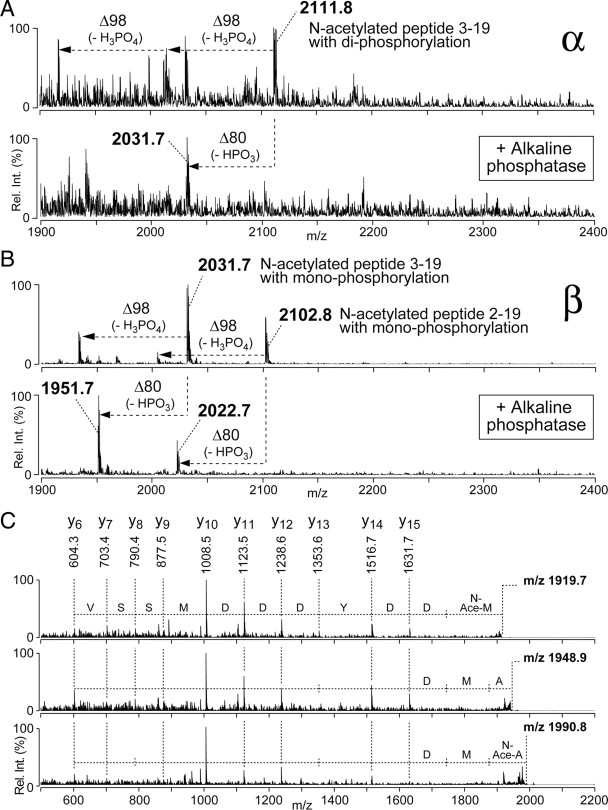
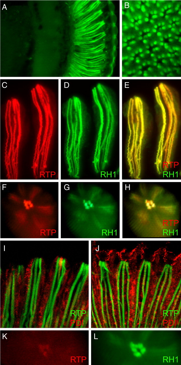
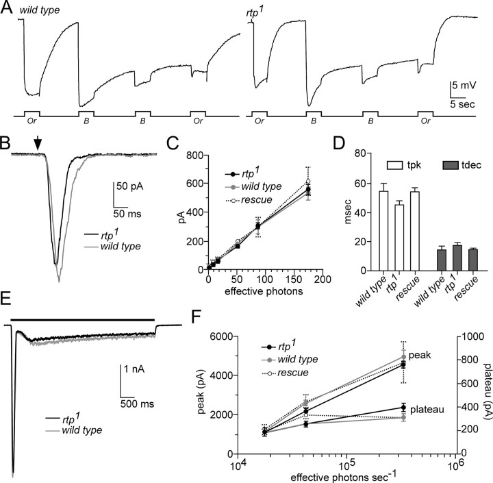
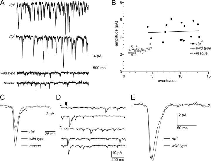
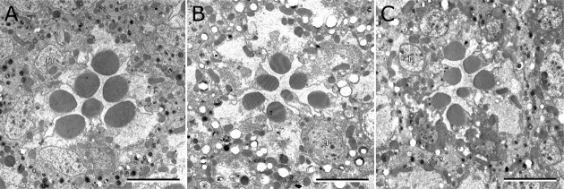
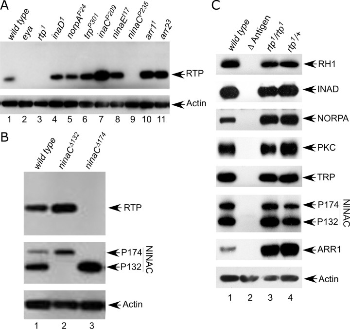
Photoreceptor cells achieve high sensitivity, reliably detecting single photons, while limiting the spontaneous activation events responsible for dark noise. We used proteomic, genetic, and electrophysiological approaches to characterize Retinophilin (RTP) (CG10233) in Drosophila photoreceptors and establish its involvement in dark-noise suppression. RTP possesses membrane occupation and recognition nexus (MORN) motifs, a structure shared with mammalian junctophilins and other membrane-associated proteins found within excitable cells. We show the MORN repeats, and both the N- and C-terminal domains, are required for RTP localization in the microvillar light-gathering organelle, the rhabdomere. RTP exists in multiple phosphorylated isoforms under dark conditions and is dephosphorylated by light exposure. An RTP deletion mutant exhibits a high rate of spontaneous membrane depolarization events in dark conditions but retains the normal kinetics of the light response. Photoreceptors lacking neither inactivation nor afterpotential C (NINAC) myosin III, a motor protein/kinase, also display a similar dark-noise phenotype as the RTP deletion. We show that NINAC mutants are depleted for RTP. These results suggest the increase in dark noise in NINAC mutants is attributable to lack of RTP and, furthermore, defines a novel role for NINAC in the rhabdomere. We propose that RTP is a light-regulated phosphoprotein that organizes rhabdomeric components to suppress random activation of the phototransduction cascade and thus increases the signaling fidelity of dark-adapted photoreceptors.


Figures

Figure 1.
Figure 1.
Identification and characterization of RTP proteins. A, The profile of retinal proteins in dark-adapted flies as analyzed by two-dimensional gel electrophoresis is shown. The boxed area identifies the location of RTP isoforms. B, The RTP region from two-dimensional profiles of retinal proteins prepared from dark- and light-adapted flies. The spots labeled α, β, γ, δ, and ε all contain the RTP protein (see Table 1). The abundance of the α spot was increased, and the δ and ε spots were decreased in the dark-adapted sample relative to the light-adapted sample. CBB, Coomassie brilliant blue. C, PMF analyses of the β spot excised from two-dimensional gel of the dark-adapted flies. The tryptic fragments labeled on the trace corresponded to peptide sequence of RTP and established that the major protein present in the β spot is RTP. The analysis was also carried on selected α, β, γ, δ, and ε spots and is summarized in Table 1. The PMF traces are shown in supplemental Figure S1 (available at www.jneurosci.org as supplemental material). D, In-gel phosphorylation analysis of the two-dimensional gel region containing RTP. The fluorescent phosphorylation sensor dye Pro-Q Diamond identified both the α and β spots in the dark-adapted profile as positive for phosphor staining. Weaker phosphor staining was seen in the light-adapted sample, and the phosphorylated α spot is greatly reduced. E, The RTP region of dark-adapted and light-adapted flies in CBB-stained two-dimensional gel (left panels) and 32P incorporation (right panels) from the analysis performed by Matsumoto and Pak (1984). The marked β spot in the dark-adapted sample showed the highest level of 32P labeling. Although the 25-year-old experiment showed more limited resolution of the spots than possible with current technology, both results show that RTP phosphorylation is reduced in the light-treated samples. F, PMF analysis of the dark-adapted β spot from the archived two-dimensional gel shown in E. The labeled tryptic fragments were derived from RTP (as identified in C), demonstrating that the major protein present in the 32P-labeled β spot was RTP.
Figure 2.
Figure 2.
Different phosphorylation states and N-terminal modifications are found in RTP isoforms. A, Enriched phosphopeptides derived from the α spot in Figure 1B were subjected to on-target alkaline phosphatase treatment using MALDI-QIT-TOF MS. The α spot contained a peptide ion at m/z 2111.8 corresponding to peptide 3–19 of RTP (top trace). Two peaks, corresponding to single and double loss of 98 Da, characteristic of the loss of one or two neutral H3PO4, were also present. After on-target alkaline phosphatase treatment (bottom trace), a dephosphorylated peptide was generated by the loss of a phosphate group (HPO3; −80 Da). B, Enriched phosphopeptides derived from the β spot in Figure 1B were subjected to on-target alkaline phosphatase treatment using MALDI-QIT-TOF MS. The β spot contained two peptide ions at m/z 2102.8 and m/z 2031.7, corresponding to RTP peptides 2–19 and 3–19, respectively (top trace), each showing single loss of 98 Da caused by loss of H3PO4. After on-target alkaline phosphatase treatment (bottom trace), a dephosphorylated peptide was generated by the loss of a phosphate group (HPO3; −80 Da). C, MS/MS analyses on the RTP N-terminal peptide ions m/z 1919.7, 1948.9, and 1990.8. The N-terminal amino acid in the m/z 1919.8 was acetylated Met3, the N-terminal amino acid in the m/z 1948.9 was unmodified Ala2, and N-terminal amino acid in m/z 1990.8 ion was acetylated Ala2.
Figure 3.
Figure 3.
RTP protein is exclusively found in the rhabdomeres of photoreceptors. A, A frontal section through the adult head shows RTP-GFP is only detected within the retina when the rtp promoter controls expression. B, A cross-section through the retina demonstrates that RTP-GFP is expressed in the rhabdomeres of all photoreceptor cells. C–H, The coexpression of RTP-RFP and RH1-GFP under pRh1-Gal4 control in photoreceptor cells R1–R6 shows RTP-RFP colocalized with RH1-GFP within the photoreceptor rhabdomere. C–E show two isolated ommatidial bundles of photoreceptors, and F–H show deep pseudopupil images taken from living flies. RFP, GFP, and merged channel images are shown. I, Photoreceptors coexpressing both GFP-RTP and PDI-RFP under pRh1-GAL4 in photoreceptor cells R1–R6 showed GFP-RTP also localized to rhabdomere and showed no overlap with cytoplasmic PDI-RFP. J, Photoreceptors coexpressing both GFP-RTP and PDI-RFP under GMR-GAL4 in all retinal cells showed only photoreceptor rhabdomeres retain RTP while PDI expression was expanded to other cell types of retina. K, pRh1-Gal4-driven expression of a ΔN-terminal RTP-RFP protein showed that RTP protein lacking the N-terminal domain was not stable in photoreceptor rhabdomere. The signal was very weak at young ages and lost by 1 d of age. L, RH1-GFP expression in the same eye displayed in K. The Rh1 rhodopsin localizes within the rhabdomere.
Figure 4.
Figure 4.
Creation and analysis of the rtp1 mutant. A, The genomic region that contains CG10233 (rtp) is flanked by the PBac insertions PBac{RB}Mms19e00210 and PBac{RB}CG12163e00209. FLP-directed recombination between these insertions produced a deletion chromosome, Df(3R)rtp1, that deleted CG10233 and disrupted the Mms19 and CG12163 genes. The 5.6 kb genomic DNA fragment containing the Mms19+ gene (Mms19 rescue construct) that rescued the zygotic lethality of Df(3R)rtp1 and the 3.7 kb genomic DNA used in the rtp rescue construct are also shown. B, Diagram showing the four deficiency chromosomes used in the analysis of the rtp gene. The location of the rtp gene in 82F6 is indicated. The rtp gene was missing in all deletions except Df(3R)5156. C, Top panels, Protein blots show that rtp/+ and rtp1/Df 5156 retain RTP protein, but RTP protein was absent in all rtp1/Df genotypes expected to lack the rtp gene. Bottom panels, Reprobing the protein blot with ARR2 antibodies demonstrated that similar levels of retinal proteins were loaded in all lanes. D, Protein blot analysis of the RTP-RFP fusion protein. The RTP-RFP 50 kDa protein was identified by both anti-RTP and anti-RFP antibodies (lanes 1–3). The levels of RTP-RFP protein was greatest when no native RTP is expressed (lane 1) but diminished in a dose-responsive manner when one copy (lane 2) or two copies (lane 3) of the native RTP were also expressed.
Figure 5.
Figure 5.
The rtp1 light response is similar to wild type. A, The electroretinogram response of white-eyed wild type (left) and the rtp1 mutant (right) were similar (n > 6). The protocol consisted of 5 s orange (Or) or blue (B) light stimuli administered in the indicated order. B, The responses of rtp1 and wild-type photoreceptors to a 1 ms test flash (arrow) containing ∼60 effective photons were similar in whole-cell recordings. C, The amplitude of the light response to increasing intensities of light stimuli was similar in rtp1 (n = 6) and wild-type photoreceptors (n = 4). The analysis of an additional control, the rtp1 mutant genotype containing an rtp+ transgene, is labeled as “rescue” (n = 4). Mean ± SEM is shown. D, Time to peak (tpk) and time to 50% decay (tdec) in rtp1 (n = 16) and the control strains (wild type, n = 5; rescue, n = 11) were similar. Mean ± SEM is shown. There was a tendency for slightly faster tpk values in rtp1, but this was not statistically significant (two-tailed t test). E, The rtp1 waveform of sustained responses to a bright (∼3 × 10⁵ effective photons s−1) 5 s light stimulus was similar to wild type. F, Statistical analysis of the amplitude of the sustained response waveform in rtp1 (n = 6), wild type (n = 5), and the rescue genotype (n = 5, except at highest intensity, n = 2). Mean ± SEM is shown. rtp1 and the control strains showed similar plateau and peak response values. All data in B–F were obtained from whole-cell recordings of photoreceptors from dissociated ommatidia, voltage clamped at −70 mV.
Figure 6.
Figure 6.
High spontaneous bump rates in the rtp1 mutant. A, A high rate of spontaneous dark events was present in the rtp1 mutant (top 2 traces from different cells) relative to both wild type (lower middle trace), and the rtp rescue genotype (bottom trace). B, Scatter plot of event rate against mean event amplitude for rtp1 cells (n = 15); wild type (n = 18) and rescue controls (n = 9). All rtp1 mutant photoreceptors are distinct from controls; regression lines show no correlation between event rate and amplitude. C, The spontaneous dark events (average waveform of >50 events in a representative cell of each genotype) in the rtp1 mutant, wild type, and the rescue genotype. Overall kinetics was similar, but event amplitude in rtp1 was approximately twice that of controls. D, Representative traces of light-induced bumps elicited by brief dim flashes containing on average approximately one effective photon (arrow) in the rtp1 mutant. The starred traces were judged to be “failures.” E, The light-induced bumps (average waveform of 30 bumps; aligned by rising phase) in rtp1 mutant and a control cell were indistinguishable. Shown are data from one cell (for additional traces and analysis, see text and supplemental Fig. S2, available at www.jneurosci.org as supplemental material).
Figure 7.
Figure 7.
Morphology of the rtp1 mutant retina. Cross-section of wild type (A), rtp1 mutant (B) photoreceptors at <1 d after eclosion and rtp1 mutant photoreceptors at 7 d (C) are shown. The sections are at comparable depths, indicated by the presence of R1–R6 photoreceptor nuclei (n) in all images. The rtp+ genotype was rtp1/+; the rtp mutant genotype was rtp1/Df(3R)5142. Scale bars, 5 μm.
Figure 8.
Figure 8.
RTP expression in mutants lacking other rhabdomeric components and rhabdomeric protein expression in the rtp1 mutant. A, The RTP protein expression was absent in ninaCP235, but was expressed in other phototransduction proteins found in the rhabdomere. Top panel, The first three lanes at left: wild type, eyes absent (eya1). eya1 and rtp1 are controls for antibody specificity. All mutants were homozygotes. The apparent increased expression of RTP in inaCP209 was not confirmed in subsequent experiments. Bottom panel, The blot was reprobed with anti-actin antibody as a protein loading control. B, RTP expression was dependent on activity of the NINAC P174 rhabdomeric protein. Top panel, Flies lacking only the NINAC P132 cytoplasmic form (lane 2) expressed RTP, but flies lacking only the NINA P174 rhabdomeric form (lane 3) did not express RTP. Middle panel, The blot was reprobed with NINAC antibodies to confirm identity of the ninaC Δ132 and ninaCΔ174 strains. Bottom panel, The blot was reprobed with anti-actin antibody as a protein loading control. C, RTP protein was not necessary for stable expression of other phototransduction proteins. The mutant alleles described in A were used as antigen-null (Δ Antigen) controls.

Similar articles

Cited by

References

    1. Ahmad ST, Natochin M, Artemyev NO, O'Tousa JE. The Drosophila rhodopsin cytoplasmic tail domain is required for maintenance of rhabdomere structure. FASEB J. 2007;21:449–455. - PubMed
    1. Arbeitman MN, Furlong EE, Imam F, Johnson E, Null BH, Baker BS, Krasnow MA, Scott MP, Davis RW, White KP. Gene expression during the life cycle of Drosophila melanogaster. Science. 2002;297:2270–2275. - PubMed
    1. Byk T, Bar-Yaacov M, Doza YN, Minke B, Selinger Z. Regulatory arrestin cycle secures the fidelity and maintenance of the fly photoreceptor cell. Proc Natl Acad Sci U S A. 1993;90:1907–1911. - PMC - PubMed
    1. Cagan RL, Ready DF. The emergence of order in the Drosophila pupal retina. Dev Biol. 1989;136:346–362. - PubMed
    1. Chorna-Ornan I, Tzarfaty V, Ankri-Eliahoo G, Joel-Almagor T, Meyer NE, Huber A, Payre F, Minke B. Light-regulated interaction of Dmoesin with TRP and TRPL channels is required for maintenance of photoreceptors. J Cell Biol. 2005;171:143–152. - PMC - PubMed

Publication types

MeSH terms