. 2010 Jan 27;30(4):1385-94.
doi: 10.1523/JNEUROSCI.3172-09.2010.

The Sushi domains of GABAB receptors function as axonal targeting signals

Affiliations

The Sushi domains of GABAB receptors function as axonal targeting signals

Barbara Biermann et al. J Neurosci. .

Abstract

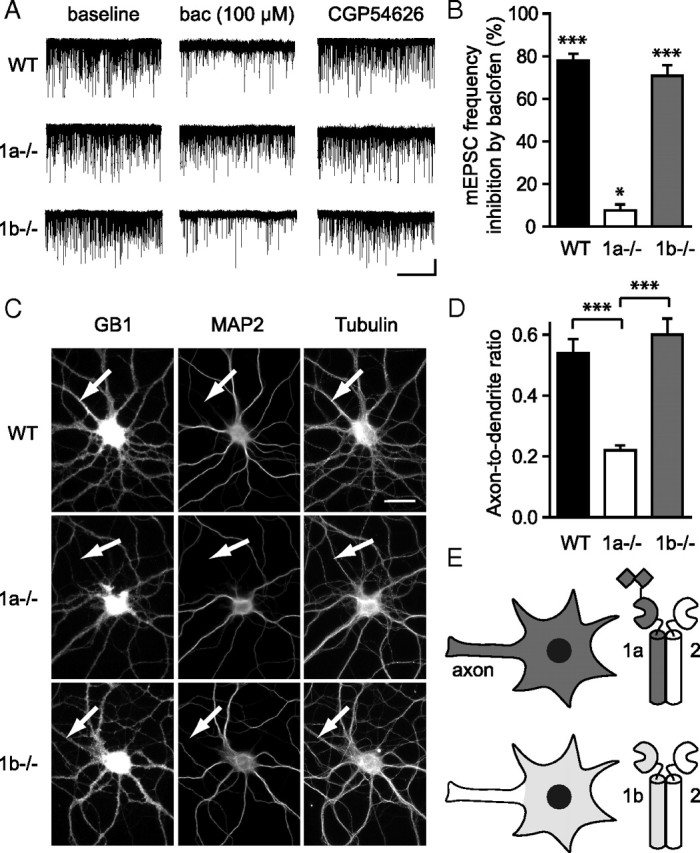
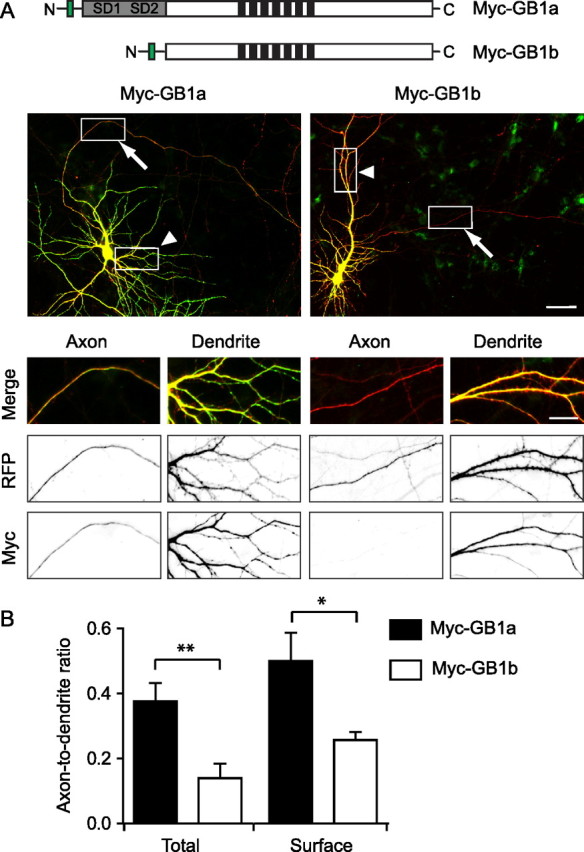
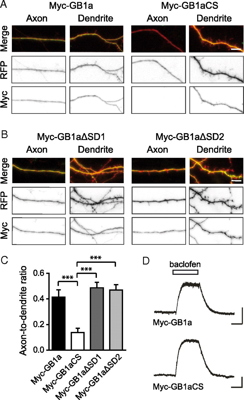
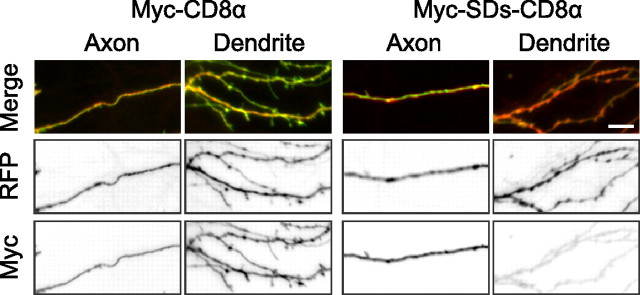
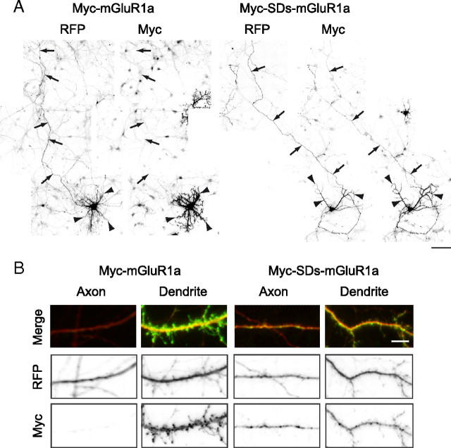
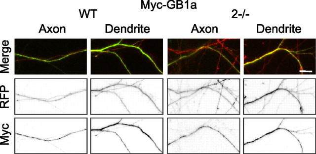
GABA(B) receptors are the G-protein-coupled receptors for GABA, the main inhibitory neurotransmitter in the brain. Two receptor subtypes, GABA(B(1a,2)) and GABA(B(1b,2)), are formed by the assembly of GABA(B1a) and GABA(B1b) subunits with GABA(B2) subunits. The GABA(B1b) subunit is a shorter isoform of the GABA(B1a) subunit lacking two N-terminal protein interaction motifs, the sushi domains. Selectively GABA(B1a) protein traffics into the axons of glutamatergic neurons, whereas both the GABA(B1a) and GABA(B1b) proteins traffic into the dendrites. The mechanism(s) and targeting signal(s) responsible for the selective trafficking of GABA(B1a) protein into axons are unknown. Here, we provide evidence that the sushi domains are axonal targeting signals that redirect GABA(B1a) protein from its default dendritic localization to axons. Specifically, we show that mutations in the sushi domains preventing protein interactions preclude axonal localization of GABA(B1a). When fused to CD8alpha, the sushi domains polarize this uniformly distributed protein to axons. Likewise, when fused to mGluR1a the sushi domains redirect this somatodendritic protein to axons, showing that the sushi domains can override dendritic targeting information in a heterologous protein. Cell surface expression of the sushi domains is not required for axonal localization of GABA(B1a). Altogether, our findings are consistent with the sushi domains functioning as axonal targeting signals by interacting with axonally bound proteins along intracellular sorting pathways. Our data provide a mechanistic explanation for the selective trafficking of GABA(B(1a,2)) receptors into axons while at the same time identifying a well defined axonal delivery module that can be used as an experimental tool.


Figures

Figure 1.
Figure 1.
Endogenous GABAB(1a,2) but not GABAB(1b,2) receptors are present in axons and inhibit glutamate release in cultured hippocampal neurons. A, Representative mEPSC recordings under baseline conditions, during baclofen (100 μm) application and after antagonizing GABAB receptors with CGP54626 (1 μm) in WT, 1a−/−, and 1b−/− neurons. Calibration: 20 pA, 25 s. B, Summary bar graph illustrating that baclofen strongly inhibits the frequency of mEPSCs in WT (78.1 ± 3.1%, n = 16) and 1b−/− (70.8 ± 5.1%, n = 15) neurons, but not in 1a−/− (7.7 ± 2.8%, n = 10) neurons. Values are means ± SEM, one-sample t test, *p < 0.05, ***p < 0.001. C, Cultured hippocampal neurons from WT, 1a−/−, and 1b−/− mice were fixed, permeabilized, and stained with antibodies recognizing GABAB1a and GABAB1b (GB1), the dendritic marker protein MAP2, or the cytoskeleton protein tubulin. Arrows mark MAP2-negative axons. Note the lack of GB1 immunolabeling in axons of 1a−/− neurons. Scale bar, 50 μm. D, A:D ratio of the endogenous GABAB1 proteins in WT, 1a−/−, and 1b−/− neurons. The fluorescence intensity of GB1 immunolabeling was normalized to the fluorescence intensity of tubulin immunolabeling. The A:D ratio of GABAB1 protein is significantly smaller in 1a−/− compared to WT and 1b−/− neurons (mean ± SEM, ***p < 0.001, 1-way ANOVA with Tukey's post hoc test). E, Schematic depiction of endogenous GABAB(1a,2) and GABAB(1b,2) receptor distribution in cultured hippocampal neurons and hippocampal slice culture. Squares indicate the two in tandem arranged SDs at the N terminus of GABAB1a.
Figure 2.
Figure 2.
Exogenous GABAB1a but not GABAB1b protein localizes to the axons of transfected hippocampal neurons in culture. A, Scheme of the tagged GABAB1 isoforms (top). The gray bar indicates the two SDs (SD1, SD2) in GABAB1a, the green bar the Myc-tag, and black bars the 7 transmembrane domains. Myc-GB1a and Myc-GB1b cDNA expression constructs were individually cotransfected with a cDNA expression construct encoding soluble RFP. Neurons were fixed at DIV14, permeabilized, and stained with antibodies recognizing MAP2 (data not shown) or the Myc-tag. Low-magnification images of the merged green Myc and the RFP fluorescence are shown at the top. Higher-magnification images of the boxed regions depict axons (arrows) and dendrites (arrowheads). Scale bars: top, 50 μm; bottom, 10 μm. B, When analyzing the total Myc-GB1a and Myc-GB1b levels in transfected neurons (Total), the A:D ratio of Myc-GB1a is significantly higher than that of Myc-GB1b (mean ± SEM, **p < 0.01, Student's t test). Likewise, when analyzing Myc-GB1a and Myc-GB1b at the cell surface of neurons coexpressing exogenous GABAB2 (Surface), the A:D ratio of Myc-GB1a is significantly higher than that of Myc-GB1b (mean ± SEM, *p < 0.05, Student's t test).
Figure 3.
Figure 3.
The SDs in GABAB1a mediate axonal localization. A, In Myc-GB1aCS, the disulfide bridges in the SDs, which are critical for ligand binding (Kirkitadze and Barlow, 2001), were disrupted by mutation of cysteines to serines. Myc-GB1a and Myc-GB1aCS were individually coexpressed with RFP in cultured hippocampal neurons. Neurons were fixed at DIV14, permeabilized, and stained with antibodies recognizing MAP2 (data not shown) and the Myc-tag. Merged images of the green Myc and the RFP fluorescence are shown at the top. Note that Myc-GB1aCS is excluded from axons. Scale bar, 10 μm. B, Myc-GB1aΔSD1 and Myc-GB1aΔSD2 proteins lacking either SD1 or SD2, respectively, both localize to axons and dendrites of transfected hippocampal neurons. Merged images of the green Myc and the RFP fluorescence are shown at the top. Scale bar, 10 μm. C, The A:D ratio of Myc-GB1aCS is significantly reduced compared to that of Myc-GB1a, while no significant reduction in the A:D ratios was observed for Myc-GB1aΔSD1 and Myc-GB1aΔSD2 (mean ± SEM, ***p < 0.001, 1-way ANOVA, Tukey's post hoc test). D, Myc-GB1aCS and Myc-GB1a, when expressed together with GABAB2, activate Kir3.1/3.2 channels in transfected CHO cells to a similar extent. Calibration: 50 pA, 5 s.
Figure 4.
Figure 4.
The SDs of GABAB1a function as axonal targeting signals in the heterologous CD8α protein. In Myc-SDs-CD8α, the SDs of GABAB1a were fused to the extracellular N-terminal domain of CD8α. Myc-CD8α or Myc-SDs-CD8α were individually coexpressed with RFP in cultured hippocampal neurons. Neurons were fixed at DIV14, permeabilized, and stained with antibodies recognizing MAP2 (data not shown) or the Myc-tag. Merged images of green Myc and RFP fluorescence are shown on top. Note that Myc-SDs-CD8α is barely detectable in dendrites, but highly expressed in axons. Scale bar, 10 μm.
Figure 5.
Figure 5.
The SDs of GABAB1a redirect the somatodendritic mGluR1a protein to axons. A, In Myc-SDs-mGluR1a, the two SDs of GABAB1a were fused to the extracellular N-terminal domain of mGluR1a. Myc-mGluR1a and Myc-SDs-mGluR1a were individually coexpressed with RFP in cultured hippocampal neurons. Neurons were fixed at DIV14, permeabilized, and stained with an antibody recognizing the Myc-tag. Arrows indicate the axon, arrowheads the dendrites. Note that Myc-SDs-mGluR1a but not Myc-mGluR1a is expressed in the axon. Scale bar, 25 μm. B, Sections of axons and dendrites of neurons expressing Myc-mGluR1a and Myc-SDs-mGluR1a. Merged images of green Myc and RFP fluorescence are shown on top. Scale bar, 10 μm.
Figure 6.
Figure 6.
Surface expression is not required for axonal trafficking of GABAB1a. Axonal and dendritic sections of cultured WT or GABA −/−B2 (2−/−) hippocampal neurons expressing Myc-GB1a and RFP. Neurons were fixed at DIV14, permeabilized, and stained with antibodies recognizing MAP2 (data not shown) or the Myc-tag. Merged images of green Myc and RFP fluorescence are shown on top. Scale bar, 10 μm.

Similar articles

Cited by

References

    1. Ahmari SE, Buchanan J, Smith SJ. Assembly of presynaptic active zones from cytoplasmic transport packets. Nat Neurosci. 2000;3:445–451. - PubMed
    1. Alle H, Geiger JR. Combined analog and action potential coding in hippocampal mossy fibers. Science. 2006;311:1290–1293. - PubMed
    1. Ango F, Albani-Torregrossa S, Joly C, Robbe D, Michel JM, Pin JP, Bockaert J, Fagni L. A simple method to transfer plasmid DNA into neuronal primary cultures: functional expression of the mGlu5 receptor in cerebellar granule cells. Neuropharmacology. 1999;38:793–803. - PubMed
    1. Aoki S, Su Q, Li H, Nishikawa K, Ayukawa K, Hara Y, Namikawa K, Kiryu-Seo S, Kiyama H, Wada K. Identification of an axotomy-induced glycosylated protein, AIGP1, possibly involved in cell death triggered by endoplasmic reticulum–Golgi stress. J Neurosci. 2002;22:10751–10760. - PMC - PubMed
    1. Aridor M, Fish KN. Selective targeting of ER exit sites supports axon development. Traffic. 2009;10:1669–1684. - PMC - PubMed

Publication types

MeSH terms