. 2010 Jan 27;30(4):1463-70.
doi: 10.1523/JNEUROSCI.4558-09.2010.

Enhanced efficacy without further cleft closure: reevaluating twist as a source of agonist efficacy in AMPA receptors

Affiliations

Enhanced efficacy without further cleft closure: reevaluating twist as a source of agonist efficacy in AMPA receptors

Amanda Birdsey-Benson et al. J Neurosci. .

Abstract

AMPA receptors (AMPARs) are tetrameric ligand-gated ion channels that couple the energy of glutamate binding to the opening of a transmembrane channel. Crystallographic and electrophysiological analysis of AMPARs has suggested a coupling between (1) cleft closure in the bilobate ligand-binding domain (LBD), (2) the resulting separation of transmembrane helix attachment points across subunit dimers, and (3) agonist efficacy. In general, more efficacious agonists induce greater degrees of cleft closure and transmembrane separation than partial agonists. Several apparent violations of the cleft-closure/efficacy paradigm have emerged, although in all cases, intradimer separation remains as the driving force for channel opening. Here, we examine the structural basis of partial agonism in GluA4 AMPARs. We find that the L651V substitution enhances the relative efficacy of kainate without increasing either LBD cleft closure or transmembrane separation. Instead, the conformational change relative to the wild-type:kainate complex involves a twisting motion with the efficacy contribution opposite from that expected based on previous analyses. As a result, channel opening may involve transmembrane rearrangements with a significant rotational component. Furthermore, a two-dimensional analysis of agonist-induced GluA2 LBD motions suggests that efficacy is not a linearly varying function of lobe 2 displacement vectors, but is rather determined by specific conformational requirements of the transmembrane domains.


Figures

Figure 1.
Figure 1.
The role of Leu651 in restricting partial-agonist-mediated cleft closure. Shown is a ribbon representation of the WT GluA4 LBD bound to glutamate (stick figure). Inset, The binding pocket of the WT GluA4 LBD in complex with Glu (blue) or KA (red), following superposition of lobe 1 Cα residues. A dashed line indicates the steric clash between the kainate isopropenyl group and the Leu651 side chain in the fully closed, glutamate-bound conformation (0.87 Å). The reorientation of lobe 2 in the kainate-bound conformation opens the cleft and relieves the steric clash (4.52 Å).
Figure 2.
Figure 2.
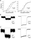
The L651V mutation increases the relative efficacy of kainate. A, Normalized whole-cell responses to kainate (left) and glutamate (right) were measured using tsA201 cells expressing GluA4 homotetramers carrying the nondesensitizing L484Y background mutation and either Leu (●) or Val (□) at position 651, as a function of agonist concentration (n = 6, kainate; n = 8, glutamate). Curves were fit using the Hill equation. B, C, Outside-out patches obtained from cells expressing GluA4-L484Y homotetramers with either Leu (B) or Val (C) at position 651 were exposed to 100 ms pulses (horizontal bars) of 10 mm KA (left) or 10 mm Glu (right), and current responses were measured under voltage-clamp conditions. Representative current recordings are shown. Note that the KA-induced currents (left) are shown at 10× scale relative to the corresponding Glu (right) responses. Averaged peak KA responses were 2.7 ± 0.4% of peak glutamate responses for channels bearing the WT Leu651 side chain (n = 5) compared with 9.4 ± 1.7% for channels bearing the Val651 mutation (n = 7).
Figure 3.
Figure 3.
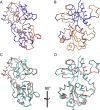
The L651V mutation permits LBD interlobe twist, but no additional cleft closure. A, B, Cα traces are shown comparing the glutamate-bound conformations of the GluA4-WT (blue) and GluA4-L651V (gold) LBD structures. The cleft is seen in side (A) and front (B) views. C, D, Corresponding side (C) and front (D) views are shown for the Cα traces of the kainate-bound GluA4-WT (cyan) and GluA4-L651V (red) LBD structures. The sideways displacement of lobe 2 between the GluA4-WT:KA and GluA4-L651V:KA structures is indicated by the arrowhead (D). In each case, superpositions were performed using lobe 1 residues as described in Materials and Methods.
Figure 4.
Figure 4.
Principal-axis analysis reveals different conformational responses of GluA4-L651V and GluA2-L650T mutants. A, B, The structure of the kainate-bound GluA4-WT LBD was aligned to its principal axes as shown in face (A) and bottom (B) views of the domain. Changes along the first (z) and second (y) principal axes primarily reflect motions associated with cleft closure, whereas changes along the third principal axis (x) reflect orthogonal motions. C, D, To visualize conformational changes associated with differences in relative efficacy, the center-of-mass displacements of lobe 2 of GluA4 (C) and GluA2 (D) LBD:agonist complexes are shown in projection along the bottom view corresponding to B, following superposition of lobe 1. In each case the WT:KA complex is chosen as the origin. Note that displacements are not on the same scale as in A and B. C, The center of mass of lobe 2 of the GluA4-WT:Glu complex (dark blue) is displaced from the GluA4-WT:KA complex (light blue) primarily along the vertical second principal axis (y), whereas the GluA4-L651V:KA complex (red) is displaced along an orthogonal pathway close to the third principal axis (x). D, The centers of mass of lobe 2 of the GluA2-WT:Glu complex (brown) and the GluA2-L650T:KA complex (purple) are both displaced from the GluA2-WT:KA complex (green) by a vertical shift primarily along the second cleft-closure axis (y).
Figure 5.
Figure 5.
Stereochemical basis for L651V-induced LBD twist. A, Following superposition of lobe 1 of the GluA4-WT:KA (cyan, gray) and GluA4-L651V:KA (red) complexes, side chains of the ligand-binding site are shown, together with the bound kainate ligand. Facilitated by a reorientation of the terminal methyl groups, the Cβ of the mutant Val651 side chain (red) occupies the position of Cγ of the WT Leu651 side chain (cyan) shifting lobe 2 sideways (arrow). B, In silico replacement of the mutant Val residue (red) of the twisted GluA4-L651V:KA complex with Leu (blue) reveals steric clashes that would occur if the WT:KA complex twisted in the same manner. These include interactions of the Cδ1 atom with the isopropenyl group of kainate (2.4 Å) and of the Cδ2 atom with atoms of Thr684, Arg685, and Thr686 (2.2–3.0 Å). C, Superposition of lobe 1 of the kainate-bound complexes of GluA2-L650T (purple) and GluA4-L651V (GluA4 in red; kainate colored by atom type) shows the horizontal displacement of the GluA4-L651V complex compared with the isosteric GluA2-L650T mutant (arrow). The hydrogen bond between Thr650 and Tyr702 in the GluA2-L650T structure is shown as a dashed line.
Figure 6.
Figure 6.
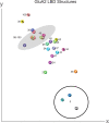
Nonlinear dependence of efficacy on cleft-closure and twist. As in Figure 4, the coordinates of lobe 1 of structures of GluA2 ligand complexes were individually aligned to those of the GluA2-WT:KA complex (1FW0) in principal-axis space. Spheres representing the center of mass of lobe 2 for each complex are shown in projection along the first principal axis with associated relative efficacy estimates (shown as percentage). Gray shading indicates a cluster of complexes showing near-maximal efficacy (98–100%). The open circle shows the position of antagonist complexes. The center-of-mass displacements are oriented primarily along a single axis, but with significant orthogonal displacements. Efficacy does not vary smoothly or monotonically in either direction. The PDB entries used for the comparison (chain A, unless otherwise indicated) are indicated by single-letter codes: A, 2CMO chain B; B, 1P1U; C, 1P1W; D, 1P1O; E, 1LBC; F, 1NNK; G, 1FTM; H, 1MM7; I, 1MQD; J, 1M5B; K, 1FTJ; L, 1M5E; M, 2AIX; N, 1SYI; O, 1MQJ; P, 1M5C; Q, 1MQI; R, 1MQH; S, 1MQG; T, 1SYH; U, 1XHY; V, 1P1N; W, 1FW0; X, 1P1U chain B; Y, 1NOT; Z, 1LB9; AA, 1FTO.

References

    1. Alam A, Jiang Y. High-resolution structure of the open NaK channel. Nat Struct Mol Biol. 2009;16:30–34. - PMC - PubMed
    1. Armstrong N, Gouaux E. Mechanisms for activation and antagonism of an AMPA-sensitive glutamate receptor: crystal structures of the GluR2 ligand binding core. Neuron. 2000;28:165–181. - PubMed
    1. Armstrong N, Mayer M, Gouaux E. Tuning activation of the AMPA-sensitive GluR2 ion channel by genetic adjustment of agonist-induced conformational changes. Proc Natl Acad Sci U S A. 2003;100:5736–5741. - PMC - PubMed
    1. Bjerrum EJ, Biggin PC. Rigid body essential X-ray crystallography: distinguishing the bend and twist of glutamate receptor ligand binding domains. Proteins. 2008;72:434–446. - PubMed
    1. Bocquet N, Nury H, Baaden M, Le Poupon C, Changeux JP, Delarue M, Corringer PJ. X-ray structure of a pentameric ligand-gated ion channel in an apparently open conformation. Nature. 2009;457:111–114. - PubMed

Publication types

MeSH terms