Comparative Study
. 2010 Jan 27;30(4):1566-74.
doi: 10.1523/JNEUROSCI.3690-09.2010.

Identification of the zebrafish ventral habenula as a homolog of the mammalian lateral habenula

Affiliations
Comparative Study

Identification of the zebrafish ventral habenula as a homolog of the mammalian lateral habenula

Ryunosuke Amo et al. J Neurosci. .

Abstract

The mammalian habenula consists of the medial and lateral habenulae. Recent behavioral and electrophysiological studies suggested that the lateral habenula plays a pivotal role in controlling motor and cognitive behaviors by influencing the activity of dopaminergic and serotonergic neurons. Despite the functional significance, manipulating neural activity in this pathway remains difficult because of the absence of a genetically accessible animal model such as zebrafish. To address the level of lateral habenula conservation in zebrafish, we applied the tract-tracing technique to GFP (green fluorescent protein)-expressing transgenic zebrafish to identify habenular neurons that project to the raphe nuclei, a major target of the mammalian lateral habenula. Axonal tracing in live and fixed fish showed projection of zebrafish ventral habenula axons to the ventral part of the median raphe, but not to the interpeduncular nucleus where the dorsal habenula projected. The ventral habenula expressed protocadherin 10a, a specific marker of the rat lateral habenula, whereas the dorsal habenula showed no such expression. Gene expression analyses revealed that the ventromedially positioned ventral habenula in the adult originated from the region of primordium lateral to the dorsal habenula during development. This suggested that zebrafish habenulae emerge during development with mediolateral orientation similar to that of the mammalian medial and lateral habenulae. These findings indicated that the lateral habenular pathways are evolutionarily conserved pathways and might control adaptive behaviors in vertebrates through the regulation of monoaminergic activities.


Figures

Figure 1.
Figure 1.
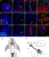
Neurons that project to the median raphe are distributed specifically in the ventral habenula. A, Sagittal section of an adult Tg(brn3a-hsp70:GFP) showing the habenulo-interpeduncular projection (green). The position of the coronal sections used in the following panels are indicated by oblique vertical bars. B, Coronal section of an adult wild-type brain showing the distributions of serotonin-positive neurons in the median raphe (green). C, D, Retrograde labeling of neurons in the ventral habenula that project to the median raphe. Coronal sections of an adult Tg(brn3a-hsp70:GFP) zebrafish showing the DiI injection site in the median raphe (C, asterisk) and the retrogradely labeled cell bodies in the ventral habenula (D, red, bracket). The GFP signal (green) marks the medial subnucleus of the dorsal habenula that projects to the IPN. E–H, Retrograde labeling of neurons in the ventral habenula that project to the median raphe (black). Coronal sections of an adult Tg(brn3a-hsp70:GFP) zebrafish showing the Neurobiotin injection site (E, asterisk), the distribution of retrogradely labeled neurons in the ventral habenula (F, brackets), sections of the fasciculi retroflexus (G, the outer sheath indicated by arrows and the core indicated by arrowhead), and the labeled fibers that pass laterally through the GFP-positive IPN (H, arrows). I, J, Coronal sections of an adult mouse brain (I) and Tg(brn3a-hsp70:GFP) zebrafish (J) showing murine Brn3a mRNA expression in the medial and lateral habenulae (I) and zebrafish brn3a mRNA (red) in the dorsal and ventral habenulae (J). K, Coronal section of an adult zebrafish habenula showing Hu immunoreactivity. L, Schematic diagram of a coronal section of zebrafish habenula showing the dorsal (white and green) and the ventral habenulae (red). The dots indicate the regions rich in cell bodies. The remaining gaps are neuropils. Sections were counterstained with fluorescent Nissl (B, K) or DAPI (J). Cbll, Cerebellum; Hb, habenula; IL, inferior lobe of the hypothalamus; IPN, interpeduncular nucleus; LHb, lateral habenula; MHb, medial habenula; Tel, telencephalon; TeO, optic tectum. Scale bars: A, 500 μm; B, C, E–H, 100 μm; D, J, K, 50 μm; I, 200 μm.
Figure 2.
Figure 2.
Ventral habenular neurons project to the ventral part of the median raphe. A–L, Anterograde tract tracing from the ventral habenula to the median raphe using Neurobiotin (A–D, red), transfected Lyn-RFP/Syp-GFP (E–H, red and green, respectively), and transfected Lyn-GFP (I–L, green). Coronal sections of the adult zebrafish showing the labeled cell bodies (A, E, I, arrows), labeled axons that circumscribe the IPN (B, F, J, arrows), the axons terminated in the ventral part of the median raphe (C, G, K, arrows), and the axonal terminals (D, H, arrowheads) in the median raphe including serotonin-positive neurons (C, green; K, L, red). The insets in E and I show higher magnification of the regions indicated by arrows in E and I, respectively. D, H, and L show higher magnification of the regions indicated by arrows in C, G, and K, respectively. The arrowheads in K and L indicate the serotonin-positive neurons. Sections were counterstained with DAPI (blue). M, N, Schematic diagram showing the dorsal oblique (M) and lateral (N) views of the axonal projections from the dorsal (red and green) and ventral habenulae (blue) in the adult zebrafish brain. The left and right dorsal habenulae are subdivided into lateral (red) and medial (green) subnuclei and show asymmetry in subnuclear size. d, Dorsal; l, left; MR, median raphe; OB, olfactory bulb; P, pineal organ; PP, parapineal organ; r, right; v, ventral. Scale bars: A–C, E–G, I–K, 50 μm; D, H, 10 μm; L, 25 μm.
Figure 3.
Figure 3.
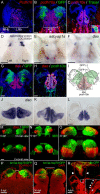
Evolutionary conservation of molecular profiles between the fish ventral habenula and mammalian lateral habenula. A, B, Coronal sections of an adult rat (A) and an adult Tg(brn3a-hsp70:GFP) zebrafish (B) showing the expression patterns of rat Pcdh10 in the lateral habenula (A, red) and zebrafish pcdh10a in the ventral habenula (B, red). vHb, Ventral habenula. The green coloration in B represents brn3a-hsp70:GFP transgene expression in the medial subnucleus of the dorsal habenula. The sections were stained with anti-HuC/D antibody (A, pseudocolored blue) or DAPI (B, blue). C, Retrograde labeling of neurons that project to the median raphe. A coronal section of wild-type zebrafish showing Neurobiotin (green), pcdh10a (red), and nuclear staining (DAPI; blue). The inset shows higher magnification of a retrogradely labeled neuron (green) coexpressing pcdh10a (yellow). D–F, GeneChip analysis identified genes specifically expressed in the zebrafish habenula. Dorsal views of the 4 dpf wild-type zebrafish brain showing the expression of hypothetical protein LOC335834 (D), adenylate cyclase activating polypeptide 1a (E), and diamine oxidase (F). G, H, Coronal sections of an adult Tg(brn3a-hsp70:GFP) (G) and wild-type (H) zebrafish localizing the expression of dao (red), brn3a-hsp70:GFP (G, green), and pcdh10a (H, green), as well as nuclear staining with DAPI (blue). I, Schematic diagram of a coronal section of adult zebrafish habenula, summarizing the expression patterns of those genes expressed specifically in the habenular subregions. J–L, Serial coronal sections of the zebrafish wild-type brain at the levels of the rostral (J), middle (K), and caudal (L) regions of the ventral habenula, showing the expression of dao. M–O, Development of the ventral habenula, as revealed by dao expression patterns. Dorsal (top panels) and coronal (bottom panels) views of the Tg(brn3a-hsp70:GFP) zebrafish showing the distributions of dao-expressing cells in the ventral habenula (red) and GFP-expressing cells in the medial subnucleus of the dorsal habenula (green). Developmental stages are shown in the left bottom corner of the top panels. P–R, Diencephalic roof remained in contact with the medial margin of the dorsal habenula throughout development. Coronal sections of the 5 dpf (P) and 30 dpf (Q) Tg(brn3a-hsp70:GFP) and adult Tg(flh:GFP) zebrafish (R) showing roofplate tela (P, Q, brackets) contacting the GFP-expressing medial subnucleus of the dorsal habenula (P, Q, green), pineal organ (R, green), and saccus dorsalis (presumptive derivative of the embryonic roofplate in the adult brain; R, arrowheads). The inset is a higher magnification of the boxed area in R showing the saccus dorsalis (arrowheads) attached to the medial margin of the habenula (asterisk). Sections were counterstained with TO-PRO-3 (P, R, red) or DAPI (Q, red). Scale bars: A, R, 100 μm; B–H, J–L, Q, 50 μm; M–P, 25 μm.
Figure 4.
Figure 4.
Medial- and lateral-habenula homologs in vertebrates. Schematic drawings of coronal sections at the level of the habenula, showing regions homologous to the mammalian medial (light green) and lateral habenulae (pink). The circles (A, B) and small dots (C–E) indicate the regions rich in cell bodies. The remaining gaps are neuropils. A–C, Schematics with modifications based on Figure 3P (A), Figure 3Q (B), and Figure 3, G and R (C) showing coronal sections of the 5 dpf (A), 30 dpf (B), and adult (C) zebrafish habenula. The red arrows in A and B indicate the morphogenetic movements of the ventral habenular primordium from the lateral to ventromedial position within the habenula according to the development. The brackets (A–C) indicate the roofplate (A) and saccus dorsalis (B, C), respectively. The red and blue lines indicate the pia mater and ventricular surface, respectively. D, Schematic with modification based on Figure 3A showing a coronal section of the rat habenula. E, Schematic modified from data presented previously by Mikula et al. (2007) showing a coronal section of the macaque monkey habenula.

Similar articles

Cited by

References

    1. Aizawa H, Bianco IH, Hamaoka T, Miyashita T, Uemura O, Concha ML, Russell C, Wilson SW, Okamoto H. Laterotopic representation of left-right information onto the dorso-ventral axis of a zebrafish midbrain target nucleus. Curr Biol. 2005;15:238–243. - PMC - PubMed
    1. Aizawa H, Goto M, Sato T, Okamoto H. Temporally regulated asymmetric neurogenesis causes left-right difference in the zebrafish habenular structures. Dev Cell. 2007;12:87–98. - PubMed
    1. Allen Institute for Brain Science. Seattle: Allen Institute for Brain Science; 2009. Allen mouse brain atlas. Available at http://mouse.brain-map.org.
    1. Almeida AP, Beaven MA. Phylogeny of histamine in vertebrate brain. Brain Res. 1981;208:244–250. - PubMed
    1. Amat J, Sparks PD, Matus-Amat P, Griggs J, Watkins LR, Maier SF. The role of the habenular complex in the elevation of dorsal raphe nucleus serotonin and the changes in the behavioral responses produced by uncontrollable stress. Brain Res. 2001;917:118–126. - PubMed

Publication types

MeSH terms

LinkOut - more resources