Comparative Study
. 2010 Apr;103(4):1856-64.
doi: 10.1152/jn.00896.2009. Epub 2010 Jan 27.

Receptive field mosaics of retinal ganglion cells are established without visual experience

Affiliations
Comparative Study

Receptive field mosaics of retinal ganglion cells are established without visual experience

Anastacia Anishchenko et al. J Neurophysiol. 2010 Apr.

Abstract

A characteristic feature of adult retina is mosaic organization: a spatial arrangement of cells of each morphological and functional type that produces uniform sampling of visual space. How the mosaics of visual receptive fields emerge in the retina during development is not fully understood. Here we use a large-scale multielectrode array to determine the mosaic organization of retinal ganglion cells (RGCs) in rats around the time of eye opening and in the adult. At the time of eye opening, we were able to reliably distinguish two types of ON RGCs and two types of OFF RGCs in rat retina based on their light response and intrinsic firing properties. Although the light responses of individual cells were not yet mature at this age, each of the identified functional RGC types formed a receptive field mosaic, where the spacing of the receptive field centers and the overlap of the receptive field extents were similar to those observed in the retinas of adult rats. These findings suggest that, although the light response properties of RGCs may need vision to reach full maturity, extensive visual experience is not required for individual RGC types to form a regular sensory map of visual space.


Figures
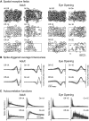
Fig. 1.
Fig. 1.
Two ON and two OFF retinal ganglion cell (RGC) types were most readily identified in adult rats and at the age of eye opening. Shown are the results from 1 adult and 1 P12 rat. A: top: spatial RFs of all OFF cells and all ON cells identified simultaneously in the same retina. Middle and Bottom: spatial RFs of the 2 ON and 2 OFF RGC types that were reliably distinguished across preparations, separately in each age group. Overlaid is a rectangular outline of the multielectrode array. B: overlaid spike-triggered average (STA) time courses for all cells of each identified RGC type. The vertical axis is normalized for each cell so that the area under each STA curve equals 1. A higher degree of the response transience is indicated by a time course with more symmetric positive and negative deflections. A 0 crossing closer to the time of spike (time 0) indicates faster response kinetics (see Fig. 2 and Table 1 for quantitative cross-type comparisons). C: overlaid autocorrelation functions for all cells of each identified RGC type. The vertical axis is normalized for each cell so that the area under each autocorrelation curve equals 1. The difference in the shape of autocorrelation functions between cell types reflects differences in intrinsic firing properties.
Fig. 2.
Fig. 2.
Comparison of RGC types within the same retina and across rats of the same age. A: comparison of light response and firing properties of RGCs belonging to different functional types. Shown are the results from 1 adult and 1 P12 rat (same animals as in Fig. 1). Each RGC type forms a cluster in the properties space. In practice, principal component analysis (PCA) was performed on the cell properties and the classification was carried out in the principal components space, which allowed for a better cluster separation. B: comparison of receptive field (RF) radius and firing properties across cell types in 3 adult rats and 3 rats at the age of eye opening (P12–P14). Within each retina, all values were normalized to the value for type OFF-1 (green) to emphasize the relative differences. Within each of the adult retinas, all pairwise differences between cell types were significant, except the difference in RF size between OFF-1A (green) and OFF-2A (red) for any of the 3 rats, and the difference between the same types in the autocorrelation steady rate for rat 1. At eye opening, all pairwise differences were significant except the difference in the autocorrelation steady rate between OFF 2E (red) and ON-2E (magenta) in rat 1 and between ON-1E (blue) and ON-2E (magenta) in rats 2 and 3. Error bars are ±SD. Note: Although the same set of colors was used to represent cell types in the adult and juvenile rats, we do not have evidence that the 4 types detected at eye opening (OFF-1E, OFF-2E, ON-1E, and ON-2E) are the same cell types that develop into the 4 types that we detected in the adult (OFF-1A, OFF-2A, ON-1A, and ON-2A).
Fig. 3.
Fig. 3.
Mosaic properties of juvenile retinas with no visual experience are comparable to the properties of mature mosaics. A: the density recovery profile shows a normalized spatial autocorrelogram of RF centers (open bars). Dotted horizontal line represents uniform cell density that would result from a random distribution. Overlaid in gray is a distribution of the nearest neighbor distances. Arrowheads mark a typical distance between the nearest neighbor RFs computed as a mean of the densest 70% of the distribution (Table 1). B: the distribution of normalized nearest neighbor distances in the units of RF radius. Arrowheads mark a typical normalized nearest neighbor distance (NND) as a mean of the densest 70% of the distribution (Table 1). For ON-2E, an asterisk above the last bin indicates that all values of NND > 5 have been also included into that bin. In both A and B, the results are from 1 adult and 1 P12 rat (same animals as in Figs. 1 and 2A).
Fig. 4.
Fig. 4.
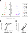
Light response kinetics are not mature at the age of eye opening. A: comparison of light response kinetics and transience between RGC types identified in 3 adult rats (■) and 3 rats at the age of eye opening (□). Each data point represents 1 cell type recorded in 1 animal. Cell type color codes are the same as in Fig. 2. Response transience (ranging from 0 to 1), polarity ([1 for ON cells, −1 for OFF cells), and time to 0 are determined from the STA time course for each cell and averaged across cells of the same type. Error bars represent SD across all cells of a given type within 1 retina. B: cumulative histograms comparing light response kinetics across all recorded RGCs. Three adult rats, dark gray; 3 rats at eye opening, light gray. Shading shows ±SD. C: same as B for light response transience.

Similar articles

Cited by

References

    1. Akerman CJ, Smyth D, Thompson ID. Visual experience before eye-opening and the development of the retinogeniculate pathway. Neuron 36: 869–879, 2002 - PubMed
    1. Bansal A, Singer JH, Hwang BJ, Xu W, Beaudet A, Feller MB. Mice lacking specific nicotinic acetylcholine receptor subunits exhibit dramatically altered spontaneous activity patterns and reveal a limited role for retinal waves in forming ON and OFF circuits in the inner retina. J Neurosci 20: 7672–7681, 2000 - PMC - PubMed
    1. Bayley PR, Morgans CW. Rod bipolar cells and horizontal cells form displaced synaptic contacts with rods in the outer nuclear layer of the nob2 retina. J Comp Neurol 500: 286–298, 2007 - PMC - PubMed
    1. Bowe-Anders C, Miller RF, Dacheux R. Developmental characteristics of receptive organization in the isolated retina-eyecup of the rabbit. Brain Res 87: 61–65, 1975 - PubMed
    1. Carcieri SM, Jacobs AL, Nirenberg S. Classification of retinal ganglion cells: a statistical approach. J Neurophysiol 90: 1704–1713, 2003 - PubMed

Publication types