. 2010 Mar 1;184(5):2611-2619.
doi: 10.4049/jimmunol.0903526. Epub 2010 Jan 27.

Anti-inflammatory role of the murine formyl-peptide receptor 2: ligand-specific effects on leukocyte responses and experimental inflammation

Affiliations

Anti-inflammatory role of the murine formyl-peptide receptor 2: ligand-specific effects on leukocyte responses and experimental inflammation

Neil Dufton et al. J Immunol. .

Erratum in

  • J Immunol. 2011 Feb 15;186(4):2684-5

Abstract

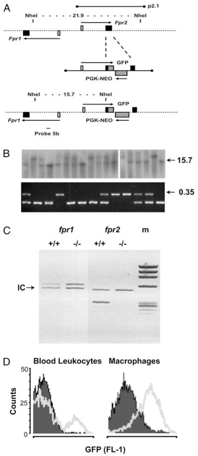
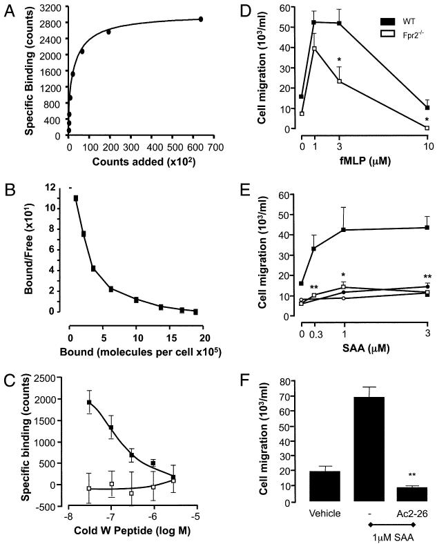
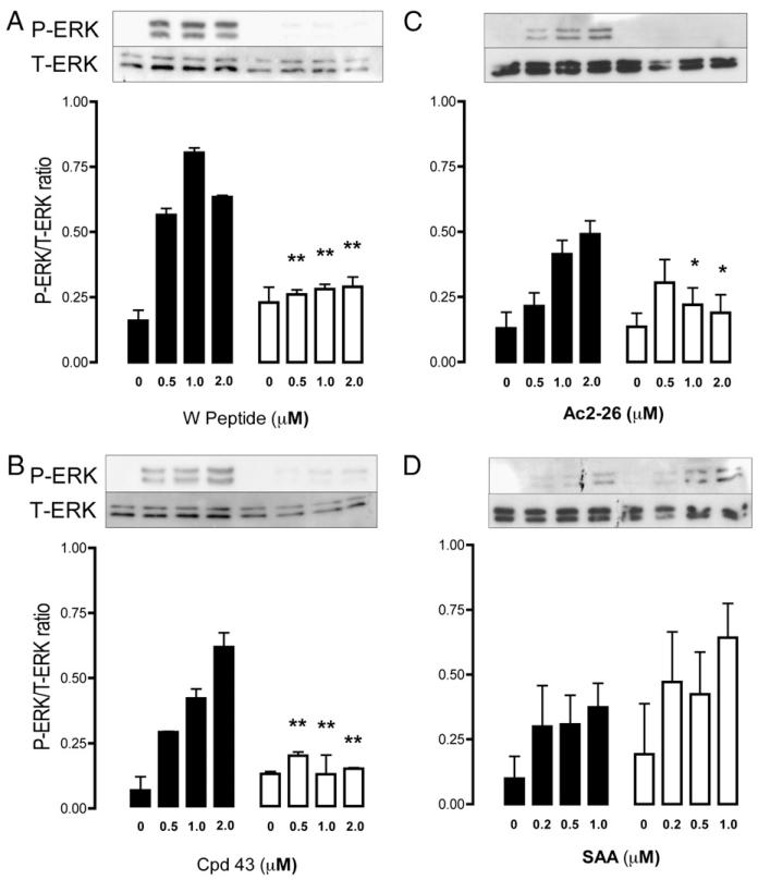
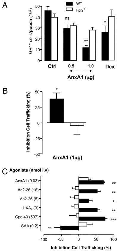
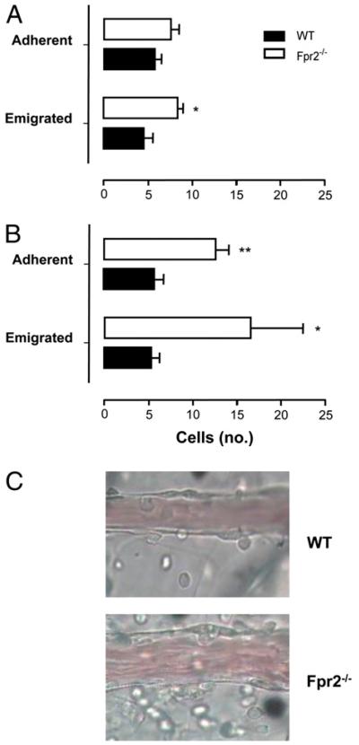
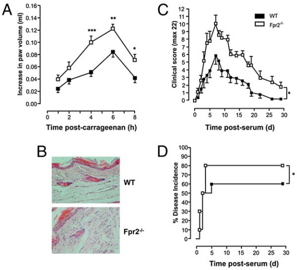
The human formyl-peptide receptor (FPR)-2 is a G protein-coupled receptor that transduces signals from lipoxin A(4), annexin A1, and serum amyloid A (SAA) to regulate inflammation. In this study, we report the creation of a novel mouse colony in which the murine FprL1 FPR2 homologue, Fpr2, has been deleted and describe its use to explore the biology of this receptor. Deletion of murine fpr2 was verified by Southern blot analysis and PCR, and the functional absence of the G protein-coupled receptor was confirmed by radioligand binding assays. In vitro, Fpr2(-/-) macrophages had a diminished response to formyl-Met-Leu-Phe itself and did not respond to SAA-induced chemotaxis. ERK phosphorylation triggered by SAA was unchanged, but that induced by the annexin A1-derived peptide Ac2-26 or other Fpr2 ligands, such as W-peptide and compound 43, was attenuated markedly. In vivo, the antimigratory properties of compound 43, lipoxin A(4), annexin A1, and dexamethasone were reduced notably in Fpr2(-/-) mice compared with those in wild-type littermates. In contrast, SAA stimulated neutrophil recruitment, but the promigratory effect was lost following Fpr2 deletion. Inflammation was more marked in Fpr2(-/-) mice, with a pronounced increase in cell adherence and emigration in the mesenteric microcirculation after an ischemia-reperfusion insult and an augmented acute response to carrageenan-induced paw edema, compared with that in wild-type controls. Finally, Fpr2(-/-) mice exhibited higher sensitivity to arthrogenic serum and were completely unable to resolve this chronic pathology. We conclude that Fpr2 is an anti-inflammatory receptor that serves varied regulatory functions during the host defense response. These data support the development of Fpr2 agonists as novel anti-inflammatory therapeutics.


Figures

FIGURE 1
FIGURE 1. Generation of the Fpr2−/− mouse colony.
A, Schematic representation of a region of ~30 kb of mouse genomic DNA spanning the fpr1 and fpr2 genes. The alignment of the 14.12-kb λ insert p2.1 is shown, along with the locations of the NheI restriction sites used for Southern blot screening. Filled boxes are coding exons; white boxes are noncoding exons; the hatched box represents the Pgk-neo cassette inserted in reverse orientation into the coding region of Fpr2; the gray box shows GFP fused in-frame with the ATG start codon; arrows indicate primary transcripts. B, Southern blot analysis (top panel) screening of tail clip DNA digested with the enzyme NheI. Probe 5b generates bands of 21.9 and 15.7 kb for WT and targeted alleles, respectively. Genotyping by multiplex PCR (bottom panel) also is reported. The F1/B11 primer pair produces a band of 233 bp using WT DNA, whereas the F1/GB4 pair gives a band of 351 bp (0.35) if the targeted allele is present. C, Multiplex PCR was used to compare the expression of fpr1 and fpr2 in WT and Fpr2−/− mice. Primers were compared with the internal control gene (18S rRNA) denoted by the arrow. D, Detection of the GFP target/reporter insert by flow cytometry. Cell samples (peripheral blood or Mϕs) from WT (opaque) or Fpr2−/− (transparent) mice as analyzed by flow cytometry in the FL-1 channel (representative of six or more distinct cell preparations).
FIGURE 2
FIGURE 2. Functional deletion of Fpr2 in vitro.
A, Specific binding of [125I]-labeled W-peptide is represented as number of molecules bound compared with the γ-counts. B, This data allowed the calculation of a Scatchard plot. C, Fpr2-specific binding to WT (black) and Fpr2−/− (white) Mϕs was assessed by measuring the competitive displacement of the [125I]-labeled W-peptide trace by cold peptide. D and E, The chemotactic responses of WT (black) and Fpr2−/− (white) Mϕs toward different concentrations of (D) fMLP, (E) SAA, or Ac2–26 were assessed by using 5-μm 96-well ChemoTx plates. Data are mean ± SEM of three experiments in quadruplicate. *p < 0.05; **p < 0.01, compared with respective WT Mϕ group by Student t test. F, WT Mϕs were pretreated with either vehicle or 1 μM Ac2–26 for 10 min prior to SAA-induced (1 μM) chemotaxis. **p < 0.01; compared with vehicle-treated group by Student t test (n = 4).
FIGURE 3
FIGURE 3. Intracellular signaling induced by Fpr2 ligation.
Phosphorylation of ERK was monitored by Western blot analysis, with Mϕs exposed to a concentration range of Wpeptide, Cpd43, Ac2–26, and SAA (A–D, respectively) for 10 min at 37°C. Representative blots are shown; respective bar histograms showing cumulative data (n = 3). Closed bars, WT Mϕs; open bars, Fpr2−/− Mϕs. Data, expressed as the ratio of phospho-ERK to total ERK, are mean ± SEM of three distinct experiments with different Mϕ cultures. *p < 0.05; **p < 0.01; compared with respective WT Mϕs by Mann-Whitney U test.
FIGURE 4
FIGURE 4. AnxA1 and other Fpr2 ligands in the air-pouch and zymosan peritonitis model.
A, AnxA1 (given −10 min) or dexamethasone (0.5 mg/kg, given −1 h) were administered i.v. prior to IL-1β (20 ng) injection into 6-d-old air pouches in WT (closed bars) and Fpr2−/− mice (open bars). *p < 0.05; **p < 0.01, significant differences between treated and untreated WT values. There was no significant difference between the values for Fpr2−/− mice with any treatment (ANOVA). B, AnxA1 (1 mg, given −10 min) was administered i.v. prior to zymosan (1 mg, i.p.) injection. Gr1+ cell influx into the air pouch or peritoneal cavity was quantified at the 4 h time point by cell counting and flow cytometry. C, Fpr2 ligands were given i.v. at the doses shown (nanomoles) with data being reported as percentage of inhibition compared with vehicle-treated mice. The IL-1β response was similar in WT and Fpr2−/− mice (~3 × 10⁶ cells per pouch). *p < 0.05; **p < 0.01; ***p < 0.001; compared with respective control values (original numbers) by Student t test. In all of the cases, data are mean ± SEM of 6–12 mice per group.
FIGURE 5
FIGURE 5. Mesenteric ischemia–reperfusion injury of WT and Fpr2−/− mice.
A and B, Mesenteric circulation was subjected to 30 min ischemia followed by (A) 45 or (B) 90 min perfusion. C, A representative field analyzed following 90 min perfusion. WT and Fpr2−/− mice, spanning 100 μm in length and surrounding 50 μm of tissue either side of the vessel wall. Data are mean ± SEM of three fields per mouse of n = 5 mice per group. **p < 0.01; *p < 0.05; compared with WT by Student t test.
FIGURE 6
FIGURE 6. Carrageenan-induced paw edema and passive serum-induced arthritis: exacerbation in Fpr2−/− mice.
Mice paws were injected with 50 μl 1% carrageenan solution. A, Time course of the paw swelling in WT and Fpr2−/− mice. Data are shown as mean ± SEM of n = 15 animals. *p < 0.05; **p < 0.01; ***p < 0.001; Student t test. B, Histological analysis in dorsal section of paws at 4 h time point. Mice received 200 μl i.p. of arthrogenic K/B × N serum. C, Time course of the clinical arthritic score in WTand Fpr2−/− mice. Data are mean ± SEM of n = 6 mice. *p < 0.05; two-way ANOVA. D, Percentage disease incidence (cut-off score ≥3). *p < 0.05; log-rank test.

Similar articles

Cited by

References

    1. Nathan C. Points of control in inflammation. Nature. 2002;420:846–852. - PubMed
    1. Serhan CN, Savill J. Resolution of inflammation: the beginning programs the end. Nat. Immunol. 2005;6:1191–1197. - PubMed
    1. Luster AD, Alon R, von Andrian UH. Immune cell migration in inflammation: present and future therapeutic targets. Nat. Immunol. 2005;6:1182–1190. - PubMed
    1. Serhan CN. Resolution phase of inflammation: novel endogenous anti-inflammatory and proresolving lipid mediators and pathways. Annu. Rev. Immunol. 2007;25:101–137. - PubMed
    1. Serhan CN, Brain SD, Buckley CD, Gilroy DW, Haslett C, O’Neill LA, Perretti M, Rossi AG, Wallace JL. Resolution of inflammation: state of the art, definitions and terms. FASEB J. 2007;21:325–332. - PMC - PubMed

Publication types

MeSH terms