. 2010 Apr 1;9(7):526-36.
doi: 10.4161/cbt.9.7.11116. Epub 2010 Apr 1.

OSU-03012 enhances Ad.7-induced GBM cell killing via ER stress and autophagy and by decreasing expression of mitochondrial protective proteins

Affiliations

OSU-03012 enhances Ad.7-induced GBM cell killing via ER stress and autophagy and by decreasing expression of mitochondrial protective proteins

Hossein A Hamed et al. Cancer Biol Ther. .

Abstract

The present studies focused on determining whether the autophagy-inducing drug OSU-03012 (AR-12) could enhance the toxicity of recombinant adenoviral delivery of melanoma differentiation associated gene-7/interleukin-24 (mda-7/IL-24) in glioblastoma multiforme (GBM) cells. The toxicity of a recombinant adenovirus to express MDA-7/IL-24 (Ad.mda-7) was enhanced by OSU-03012 in a diverse panel of primary human GBM cells. The enhanced toxicity correlated with reduced ERK1/2 phosphorylation and expression of MCL-1 and BCL-XL, and was blocked by molecular activation of ERK1/2 and by inhibition of the intrinsic, but not the extrinsic, apoptosis pathway. Both OSU-03012 and expression of MDA-7/IL-24 increased phosphorylation of PKR-like endoplasmic reticulum kinase (PERK) that correlated with increased levels of autophagy and expression of dominant negative PERK blocked autophagy induction and tumor cell death. Knockdown of ATG5 or Beclin1 suppressed OSU-03012 enhanced MDA-7/IL-24-induced autophagy and blocked the lethal interaction between the two agents. Ad.mda-7-infected GBM cells secreted MDA-7/IL-24 into the growth media and this conditioned media induced expression of MDA-7/IL-24 in uninfected GBM cells. OSU-03012 interacted with conditioned media to kill GBM cells and knockdown of MDA-7/IL-24 in these cells suppressed tumor cell killing. Collectively, our data demonstrate that the induction of autophagy and mitochondrial dysfunction by a combinatorial treatment approach represents a potentially viable strategy to kill primary human GBM cells.


Figures

Figure 1
Figure 1
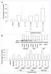
Figure 1A and B. Ad.mda-7 lethality is enhanced by OSU-03012. (A) GBM6, GBM12 and GBM14 cells were infected with empty vector control virus (Ad.cmv) or with virus to express MDA-7/IL-24 (Ad.mda-7) and 12 h after infection treated with vehicle (DMSO) or OSU-03012 (OSU, 1 μM). Forty eight h after infection cells were isolated and cell viability was determined by trypan blue exclusion assay (±SEM, n = 3). (B) GBM6 cells were plated as single cells in sextuplicate and 12 h after plating infected with Ad.cmv or Ad.mda-7 and 12 h after infection treated with vehicle (DMSO) or OSU-03012 (OSU, 1 μM). Forty eight h after infection the growth media was removed and replaced with new media lacking drugs. Colonies of >50 cells were permitted to form over the following ~20 days, followed by fixing, staining and counting (±SEM, n = 3). Figure 1C and D. Ad.mda-7 lethality is enhanced by OSU-03012. (C) GBM6 cells were infected with Ad.cmv or Ad.mda-7 and 12 h after infection treated with vehicle (DMSO) or OSU-03012 (OSU, 1 μM). Twenty four h and 48 h after infection cells were isolated and SDS PAGE performed to determine the expression of BCL-XL and MCL-1, and the phosphorylation of ERK1/2, p38 MAPK, JNK1-3, AKT (S473) (n = 2). (D) GBM6 cells were infected with Ad.cmv or Ad.mda-7 in combination with viruses to express XIAP, BCL-XL, dominant negative caspase 9 or c-FLIP-s, and 12 h after infection treated with vehicle (DMSO), or OSU-03012 (OSU, 1 μM). Forty-eight h after infection cells were isolated and cell viability was determined by trypan blue exclusion assay (±SEM, n = 3).
Figure 1
Figure 1
Figure 1A and B. Ad.mda-7 lethality is enhanced by OSU-03012. (A) GBM6, GBM12 and GBM14 cells were infected with empty vector control virus (Ad.cmv) or with virus to express MDA-7/IL-24 (Ad.mda-7) and 12 h after infection treated with vehicle (DMSO) or OSU-03012 (OSU, 1 μM). Forty eight h after infection cells were isolated and cell viability was determined by trypan blue exclusion assay (±SEM, n = 3). (B) GBM6 cells were plated as single cells in sextuplicate and 12 h after plating infected with Ad.cmv or Ad.mda-7 and 12 h after infection treated with vehicle (DMSO) or OSU-03012 (OSU, 1 μM). Forty eight h after infection the growth media was removed and replaced with new media lacking drugs. Colonies of >50 cells were permitted to form over the following ~20 days, followed by fixing, staining and counting (±SEM, n = 3). Figure 1C and D. Ad.mda-7 lethality is enhanced by OSU-03012. (C) GBM6 cells were infected with Ad.cmv or Ad.mda-7 and 12 h after infection treated with vehicle (DMSO) or OSU-03012 (OSU, 1 μM). Twenty four h and 48 h after infection cells were isolated and SDS PAGE performed to determine the expression of BCL-XL and MCL-1, and the phosphorylation of ERK1/2, p38 MAPK, JNK1-3, AKT (S473) (n = 2). (D) GBM6 cells were infected with Ad.cmv or Ad.mda-7 in combination with viruses to express XIAP, BCL-XL, dominant negative caspase 9 or c-FLIP-s, and 12 h after infection treated with vehicle (DMSO), or OSU-03012 (OSU, 1 μM). Forty-eight h after infection cells were isolated and cell viability was determined by trypan blue exclusion assay (±SEM, n = 3).
Figure 2
Figure 2
Activation of MEK and constitutive MCL-1 expression protect GBM cells from MDA-7/IL-24 and OSU-03012 toxicity. (A) GBM6 cells were infected with empty vector control virus (Ad.cmv) or with viruses to express MDA-7/IL-24 (Ad.mda-7) or a constitutively active form of MEK1 (MEK1 EE). Twelve h after infection cells were treated with vehicle (DMSO) or OSU-03012 (OSU, 1 μM). Cells were isolated 48 h after infection and cell viability was determined by trypan blue exclusion assay (±SEM, n = 3). (B) GBM6 cells were infected with empty vector control virus (Ad.cmv) or with viruses to express MDA-7/IL-24 (Ad.mda-7) and in parallel transfected with a control plasmid or a plasmid to express MCL-1. Twelve h after infection cells were treated with vehicle (DMSO) or OSU-03012 (OSU, 1 μM). Cells were isolated 48 h after infection and cell viability was determined by trypan blue exclusion assay (±SEM, n = 3).
Figure 3
Figure 3
Figure 3A and B. OSU-03012 treatment enhances Ad.mda-7-induced toxic autophagy but does not promote additional activation of PERK. (A) GBM6 cells were infected with Ad.cmv or Ad.mda-7 and 12 h after infection treated with vehicle (DMSO) or OSU-03012 (OSU, 1 μM). Cells were isolated 24 h and 48 h after infection and SDS PAGE and immunoblotting performed to assess the phosphorylation of PERK and the conversion of LC3 to LC3-II (n = 3). (B) GBM6 cells in were infected with Ad.cmv or Ad.mda-7 and in parallel transfected with either an empty vector plasmid (CMV) or a plasmid to express dominant negative PERK (dnPERK). Twelve h after infection cells were treated with vehicle (DMSO) or OSU-03012. Forty-eight h after infection cells were isolated and cell viability was determined by trypan blue dye exclusion assay (±SEM, n = 3). Figure 3C and D. OSU-03012 treatment enhances Ad.mda-7-induced toxic autophagy but does not promote additional activation of PERK. (C) GBM6 cells in 4-well chambered slides were infected with Ad.cmv or Ad.mda-7 and in parallel transfected with a plasmid to express LC3-GFP and with either an empty vector plasmid (CMV) or a plasmid to express dominant negative PERK (dnPERK). Twelve h after infection cells are treated with vehicle (DMSO) or OSU-03012. Twelve h after drug treatment cells were examined using a fluorescent microscope (×40 mag) for the formation of intense GFP staining vesicles (±SEM, n = 3). (D) GBM6 cells were infected with Ad.cmv or Ad.mda-7 and in parallel transfected with either a scrambled siRNA (siSCR) or siRNAs to knock down Beclin1 (siBeclin1) or ATG5 (siATG5). Twelve h after infection cells are treated with vehicle (DMSO) or OSU-03012 (OSU, 1 μM). Forty eight h after drug treatment cells were isolated and cell viability was determined by trypan blue exclusion assay (±SEM, n = 3).
Figure 3
Figure 3
Figure 3A and B. OSU-03012 treatment enhances Ad.mda-7-induced toxic autophagy but does not promote additional activation of PERK. (A) GBM6 cells were infected with Ad.cmv or Ad.mda-7 and 12 h after infection treated with vehicle (DMSO) or OSU-03012 (OSU, 1 μM). Cells were isolated 24 h and 48 h after infection and SDS PAGE and immunoblotting performed to assess the phosphorylation of PERK and the conversion of LC3 to LC3-II (n = 3). (B) GBM6 cells in were infected with Ad.cmv or Ad.mda-7 and in parallel transfected with either an empty vector plasmid (CMV) or a plasmid to express dominant negative PERK (dnPERK). Twelve h after infection cells were treated with vehicle (DMSO) or OSU-03012. Forty-eight h after infection cells were isolated and cell viability was determined by trypan blue dye exclusion assay (±SEM, n = 3). Figure 3C and D. OSU-03012 treatment enhances Ad.mda-7-induced toxic autophagy but does not promote additional activation of PERK. (C) GBM6 cells in 4-well chambered slides were infected with Ad.cmv or Ad.mda-7 and in parallel transfected with a plasmid to express LC3-GFP and with either an empty vector plasmid (CMV) or a plasmid to express dominant negative PERK (dnPERK). Twelve h after infection cells are treated with vehicle (DMSO) or OSU-03012. Twelve h after drug treatment cells were examined using a fluorescent microscope (×40 mag) for the formation of intense GFP staining vesicles (±SEM, n = 3). (D) GBM6 cells were infected with Ad.cmv or Ad.mda-7 and in parallel transfected with either a scrambled siRNA (siSCR) or siRNAs to knock down Beclin1 (siBeclin1) or ATG5 (siATG5). Twelve h after infection cells are treated with vehicle (DMSO) or OSU-03012 (OSU, 1 μM). Forty eight h after drug treatment cells were isolated and cell viability was determined by trypan blue exclusion assay (±SEM, n = 3).
Figure 4
Figure 4
Figure 4A and B. MDA-7/IL-24 stimulates expression of MDA-7/IL-24 in GBM cells in a PERK-dependent fashion. (A) GBM6 cells were infected with Ad.cmv or Ad.mda-7. Forty-eight h after infection cells and media were isolated. Cell viability was determined by trypan blue dye exclusion assay (±SEM, n = 3). The media was placed onto GBM6 cells and 24 h after media addition cells were irradiated (4 Gy). Forty-eight h after placement of conditioned media onto the cells, cell viability was again determined by trypan blue dye exclusion assay (±SEM, n = 3). (B) GBM6 cells were infected with Ad.cmv or Ad.mda-7. Forty-eight h after infection cells and media were isolated. Cell viability was determined by trypan blue dye exclusion assay (±SEM, n = 3). The media was placed onto GBM6 cells that had been transfected with plasmids to express a control shRNA (shSCR) or an shRNA to knock down MDA-7/IL-24 expression (shMDA-7) (labeled in parentheses, grey). Thirty min after media addition cells were treated with: (a) radiation (4 Gy) or; (b) vehicle (DMSO) or OSU-03012 (OSU, 1 μM). Forty-eight h after placement of conditioned media onto the cells, cell viability was again determined by trypan blue exclusion assay (±SEM, n = 3). Inset Panel: conditioned media containing MDA-7/IL-24 induces MDA-7/IL-24 in uninfected GBM cells that is blocked by shRNA knock down of MDA-7/IL-24. Figure 4C and D. MDA-7/IL-24 stimulates expression of MDA-7/IL-24 in GBM cells in a PERK-dependent fashion. (C) GBM6 cells were infected with Ad.cmv or Ad.mda-7. Forty-eight h after infection cells and media were isolated. Cell viability was determined by trypan blue exclusion assay (±SEM, n = 3). The media was placed onto GBM6 cells, Twenty-four h after media addition cells were treated with: vehicle (DMSO) or OSU-03012 (OSU, 1 μM). Forty-eight h after placement of conditioned media onto the cells, cell viability was again determined by trypan blue dye exclusion assay (±SEM, n = 3). (D) GBM6 cells were infected with Ad.cmv or Ad.mda-7. Forty-eight h after infection the growth media was isolated. The media was placed onto GBM6 cells that had been transfected with plasmids to express a nothing (CMV) or to express dominant negative PERK (dnPERK) (labeled in parentheses, grey). Forty-eight h after placement of conditioned media onto the cells, cell viability was again determined by trypan blue exclusion assay (±SEM, n = 3) *p < 0.05 less than corresponding value in parallel empty vector control transfected cells.
Figure 4
Figure 4
Figure 4A and B. MDA-7/IL-24 stimulates expression of MDA-7/IL-24 in GBM cells in a PERK-dependent fashion. (A) GBM6 cells were infected with Ad.cmv or Ad.mda-7. Forty-eight h after infection cells and media were isolated. Cell viability was determined by trypan blue dye exclusion assay (±SEM, n = 3). The media was placed onto GBM6 cells and 24 h after media addition cells were irradiated (4 Gy). Forty-eight h after placement of conditioned media onto the cells, cell viability was again determined by trypan blue dye exclusion assay (±SEM, n = 3). (B) GBM6 cells were infected with Ad.cmv or Ad.mda-7. Forty-eight h after infection cells and media were isolated. Cell viability was determined by trypan blue dye exclusion assay (±SEM, n = 3). The media was placed onto GBM6 cells that had been transfected with plasmids to express a control shRNA (shSCR) or an shRNA to knock down MDA-7/IL-24 expression (shMDA-7) (labeled in parentheses, grey). Thirty min after media addition cells were treated with: (a) radiation (4 Gy) or; (b) vehicle (DMSO) or OSU-03012 (OSU, 1 μM). Forty-eight h after placement of conditioned media onto the cells, cell viability was again determined by trypan blue exclusion assay (±SEM, n = 3). Inset Panel: conditioned media containing MDA-7/IL-24 induces MDA-7/IL-24 in uninfected GBM cells that is blocked by shRNA knock down of MDA-7/IL-24. Figure 4C and D. MDA-7/IL-24 stimulates expression of MDA-7/IL-24 in GBM cells in a PERK-dependent fashion. (C) GBM6 cells were infected with Ad.cmv or Ad.mda-7. Forty-eight h after infection cells and media were isolated. Cell viability was determined by trypan blue exclusion assay (±SEM, n = 3). The media was placed onto GBM6 cells, Twenty-four h after media addition cells were treated with: vehicle (DMSO) or OSU-03012 (OSU, 1 μM). Forty-eight h after placement of conditioned media onto the cells, cell viability was again determined by trypan blue dye exclusion assay (±SEM, n = 3). (D) GBM6 cells were infected with Ad.cmv or Ad.mda-7. Forty-eight h after infection the growth media was isolated. The media was placed onto GBM6 cells that had been transfected with plasmids to express a nothing (CMV) or to express dominant negative PERK (dnPERK) (labeled in parentheses, grey). Forty-eight h after placement of conditioned media onto the cells, cell viability was again determined by trypan blue exclusion assay (±SEM, n = 3) *p < 0.05 less than corresponding value in parallel empty vector control transfected cells.

Comment in

Similar articles

Cited by

References

    1. Robins HI, Chang S, Butowski N, Mehta M. Therapeutic advances for glioblastoma multiforme: current status and future prospects. Curr Oncol Rep. 2007;9:66–70. - PubMed
    1. Jiang H, Lin JJ, Su ZZ, Goldstein NI, Fisher PB. Subtraction hybridization identifies a novel melanoma differentiation associated gene, mda-7, modulated during human melanoma differentiation, growth and progression. Oncogene. 1995;11:2477–86. - PubMed
    1. Ekmekcioglu S, Ellerhorst J, Mhashilkar AM, Sahin AA, Read CM, Prieto VG, et al. Downregulated melanoma differentiation associated gene (mda-7) expression in human melanomas. Int J Cancer. 2001;94:54–9. - PubMed
    1. Ellerhorst JA, Prieto VG, Ekmekcioglu S. Loss of MDA-7 expression with progression of melanoma. J Clin Oncol. 2002;20:1069–74. - PubMed
    1. Huang EY, Madireddi MT, Gopalkrishnan RV, Leszczyniecka M, Su Z, Lebedeva IV, et al. Genomic structure, chromosomal localization and expression profile of a novel melanoma differentiation associated (mda-7) gene with cancer specific growth suppressing and apoptosis inducing properties. Oncogene. 2001;20:7051–63. - PubMed

Publication types

MeSH terms