

Mutations in a gene encoding a midbody protein in binucleated Reed-Sternberg cells of Hodgkin lymphoma
- PMID: 20107318
- PMCID: PMC4523393
- DOI: 10.4161/cc.9.4.10780
Mutations in a gene encoding a midbody protein in binucleated Reed-Sternberg cells of Hodgkin lymphoma
Abstract
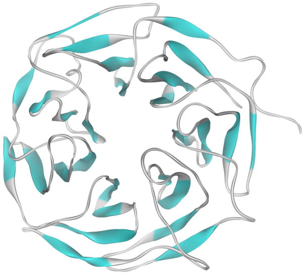
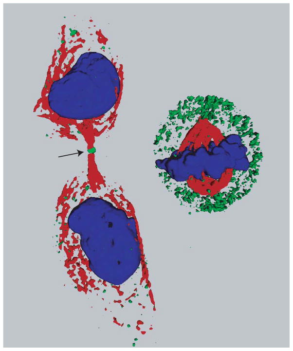
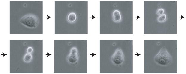
Classical Hodgkin lymphoma (cHL) is a cancer in which malignant "Reed-Sternberg" cells comprise just a fraction of the bulk of the tumor and are characteristically binucleated. We recently identified a novel gene, KLHDC8B, which appears responsible for some familial cases of cHL. KLHDC8B encodes a midbody kelch protein expressed during cytokinesis. Deficiency of KLHDC8B leads to binucleated cells, implicating its involvement in Reed-Sternberg cell formation. Interestingly, other cancer genes, such as BRCA1 and BRCA2, also encode proteins locating to the midbody during cytokinesis, even though their participation in other pathways has received greater attention. Midbody components may be an overlooked source of tumor suppressor genes.
Figures
Similar articles
-
The kelch protein KLHDC8B guards against mitotic errors, centrosomal amplification, and chromosomal instability.J Biol Chem. 2012 Nov 9;287(46):39083-93. doi: 10.1074/jbc.M112.390088. Epub 2012 Sep 17. J Biol Chem. 2012. PMID: 22988245 Free PMC article.
-
Mutations in a gene encoding a midbody kelch protein in familial and sporadic classical Hodgkin lymphoma lead to binucleated cells.Proc Natl Acad Sci U S A. 2009 Sep 1;106(35):14920-5. doi: 10.1073/pnas.0904231106. Epub 2009 Aug 12. Proc Natl Acad Sci U S A. 2009. PMID: 19706467 Free PMC article.
-
Detection of genomic imbalances in microdissected Hodgkin and Reed-Sternberg cells of classical Hodgkin's lymphoma by array-based comparative genomic hybridization.Haematologica. 2008 Sep;93(9):1318-26. doi: 10.3324/haematol.12875. Epub 2008 Jul 18. Haematologica. 2008. PMID: 18641027
-
Hodgkin lymphoma: Pathology and biology.Semin Hematol. 2016 Jul;53(3):139-47. doi: 10.1053/j.seminhematol.2016.05.007. Epub 2016 May 13. Semin Hematol. 2016. PMID: 27496304 Review.
-
The biology of classical Hodgkin lymphoma.Semin Hematol. 2024 Aug;61(4):212-220. doi: 10.1053/j.seminhematol.2024.05.001. Epub 2024 May 8. Semin Hematol. 2024. PMID: 38824068 Review.
Cited by
-
Distinct roles for TET family proteins in regulating human erythropoiesis.Blood. 2017 Apr 6;129(14):2002-2012. doi: 10.1182/blood-2016-08-736587. Epub 2017 Feb 6. Blood. 2017. PMID: 28167661 Free PMC article.
-
RNA-seq analysis identifies cytoskeletal structural genes and pathways for meat quality in beef.PLoS One. 2020 Nov 11;15(11):e0240895. doi: 10.1371/journal.pone.0240895. eCollection 2020. PLoS One. 2020. PMID: 33175867 Free PMC article.
-
The kelch protein KLHDC8B guards against mitotic errors, centrosomal amplification, and chromosomal instability.J Biol Chem. 2012 Nov 9;287(46):39083-93. doi: 10.1074/jbc.M112.390088. Epub 2012 Sep 17. J Biol Chem. 2012. PMID: 22988245 Free PMC article.
-
Common origin and somatic mutation patterns of composite lymphomas and leukemias.Leukemia. 2025 Aug;39(8):1960-1971. doi: 10.1038/s41375-025-02549-y. Epub 2025 May 22. Leukemia. 2025. PMID: 40404986 Free PMC article.
-
Incomplete cytokinesis and re-fusion of small mononucleated Hodgkin cells lead to giant multinucleated Reed-Sternberg cells.Proc Natl Acad Sci U S A. 2013 Dec 17;110(51):20729-34. doi: 10.1073/pnas.1312509110. Epub 2013 Dec 3. Proc Natl Acad Sci U S A. 2013. PMID: 24302766 Free PMC article.
References
-
- Kuppers R. The biology of Hodgkin’s lymphoma. Nat Rev Cancer. 2009;9:15–27. - PubMed
-
- Kutok JL, Wang F. Spectrum of Epstein-Barr virus-associated diseases. Annu Rev Pathol. 2006;1:375–404. - PubMed
-
- Altieri A, Hemminki K. The familial risk of Hodgkin’s lymphoma ranks among the highest in the Swedish Family-Cancer Database. Leukemia. 2006;20:2062–3. - PubMed
-
- Diepstra A, Niens M, te Meerman GJ, Poppema S, van den Berg A. Genetic susceptibility to Hodgkin’s lymphoma associated with the human leukocyte antigen region. Eur J Haematol Suppl. 2005:34–41. - PubMed
-
- Coutinho A, Carneiro-Sampaio M. Primary immunodeficiencies unravel critical aspects of the pathophysiology of autoimmunity and of the genetics of autoimmune disease. J Clin Immunol. 2008;28 (Suppl 1):S4–10. - PubMed
Publication types
MeSH terms
Substances
Grants and funding
LinkOut - more resources
Full Text Sources
Medical
Molecular Biology Databases
Miscellaneous