. 2010 Feb 25;463(7284):1035-41.
doi: 10.1038/nature08797. Epub 2010 Jan 27.

Direct conversion of fibroblasts to functional neurons by defined factors

Affiliations

Direct conversion of fibroblasts to functional neurons by defined factors

Thomas Vierbuchen et al. Nature. .

Abstract

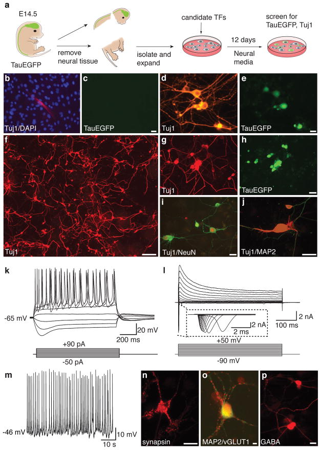
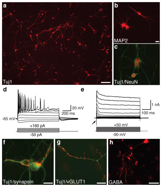
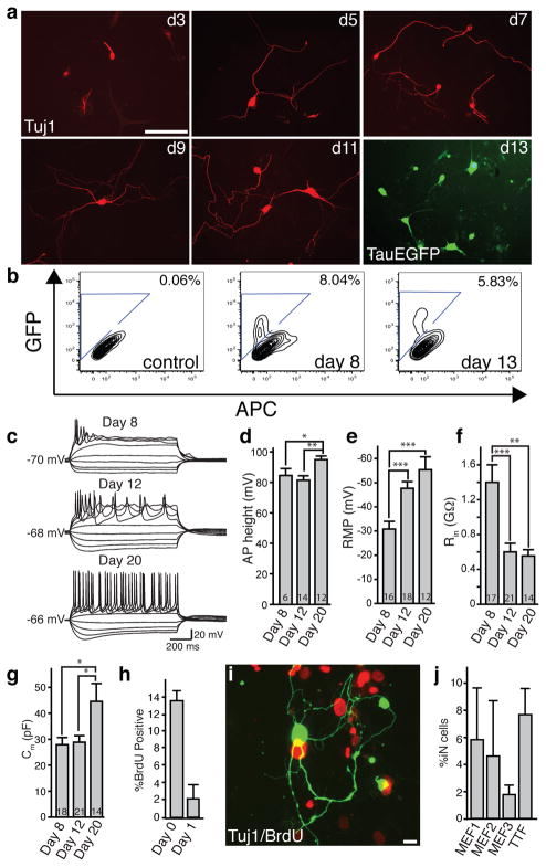
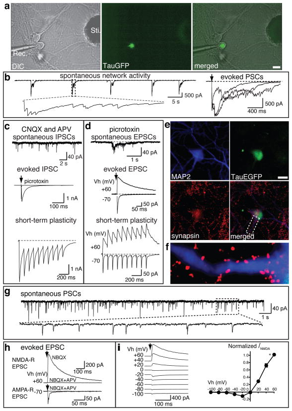
Cellular differentiation and lineage commitment are considered to be robust and irreversible processes during development. Recent work has shown that mouse and human fibroblasts can be reprogrammed to a pluripotent state with a combination of four transcription factors. This raised the question of whether transcription factors could directly induce other defined somatic cell fates, and not only an undifferentiated state. We hypothesized that combinatorial expression of neural-lineage-specific transcription factors could directly convert fibroblasts into neurons. Starting from a pool of nineteen candidate genes, we identified a combination of only three factors, Ascl1, Brn2 (also called Pou3f2) and Myt1l, that suffice to rapidly and efficiently convert mouse embryonic and postnatal fibroblasts into functional neurons in vitro. These induced neuronal (iN) cells express multiple neuron-specific proteins, generate action potentials and form functional synapses. Generation of iN cells from non-neural lineages could have important implications for studies of neural development, neurological disease modelling and regenerative medicine.


Figures

Figure 1
Figure 1. A screen for neuronal fate inducing factors and characterization of MEF-derived iN cells
a, Experimental rationale. b, Uninfected, p3 TauEGFP MEFs contained rare Tuj1-positive cells (red) with flat morphology. Blue: DAPI counterstain. c, Tuj1-positive fibroblasts do not express visible TauEGFP. d–e, MEF-iN cells express Tuj1 (red) and TauEGFP (green) and display complex neuronal morphologies 32 days after infection with the 19-factor (19F) pool. f, Tuj1 expression in MEFs 13 days after infection with the 5F pool. g–j, MEF-derived Tuj1-positive iN cells co-express the pan-neuronal markers TauEGFP (h), NeuN (red,i) and MAP2 (red,j). k, Representative traces of membrane potential responding to step depolarization by current injection (lower panel). Membrane potential was current-clamped at around −65 mV. l, Representative traces of whole-cell currents in voltage-clamp mode, cell was held at −70 mV, step depolarization from −90 mV to 60 mV at 10 mV interval were delivered (lower panel). Insert showing Na+currents. m, Spontaneous action potentials (AP) recorded from a 5F MEF-iN cell 8 days post infection. No current injection was applied. n–p, 22 days post-infection 5F MEF-iN cells expressed synapsin (red,n) and vesicular glutamate transporter 1 (vGLUT1) (red,o) or GABA (p). Scale bars = 5 μm (o), 10 μm (e,n,p) 20 μm (c,h,i), and 200 μm (f).
Figure 2
Figure 2. Efficient induction of neurons from perinatal tail-tip fibroblasts
a, Tuj1-stained tail-tip fibroblast 13 days after infection 5F pool. b–c, TTF-iNs express the pan-neuronal markers MAP2 (b) and NeuN (c). d, Representative traces showing action potentials elicited at day 13 post infection. Nine of eleven cells recorded exhibited APs. e, Whole cell currents recorded in voltage-clamp mode. Inward fast inactivating sodium currents (arrow) and outward currents can be observed. f–h, 21 days after infection TTF-iN cells express synapsin (red, f), vGLUT1 (red g) and GABA (h). c, f, and g are overlay images with the indicated marker (red) and Tuj1 (green). Scale bars = 20 μm (b,f,g), 100 μm (h), 200 μm (a).
Figure 3
Figure 3. The 5F pool induced conversion is rapid and efficient
a, Tuj1-positive iN cells (red) exhibit morphological maturation over time after viral infections. At day 13, TauEGFP expression outlines neuronal processes. b, FACS analysis of TauEGFP expression 8 and 13 days post infection. Control = Uninfected TauEGFP MEFs. c, Representative traces showing action potentials elicited from MEF-iN cells at days 8, 12, and 20 post infection. Cells were maintained at a potential of ~ −65 to −70mV. Step current injection protocols were used from −50 to +70 pA. Scale bars apply to all traces. d–g, Quantification of membrane properties in MEF-iN cells at 8, 12, and 20 days post infection. Numbers in the bars represent the numbers of recorded cells. Data are presented as mean±S.E.M. * p<0.05; **p<0.01; *** p<0.001 (Student’s t-test). AP: Action Potentials; RMP: Resting Membrane Potentials; Rin: Membrane input resistances; Cm: Membrane Capacitance. AP heights were measured from the baseline. h, BrdU-positive iN cells following BrdU treatment from day 0–13 or day 1–13 after transgene induction. i, Example of a Tuj1 (green) positive cell not labeled with BrdU (red) when added at day 0 after addition of doxycycline. Data are presented as mean ±S.D. j, Efficiency estimates for iN cell generation 13 days after infection (see methods). Every bar represents an independent experiment. Doxycycline was added to 48 hours after plating in MEF experiment #1 and after 24 hours in MEF experiments #2, #3. Error bars = ±1 S.D. of cell counts. Scale Bars = 10 μm (j), 100 μm (a).
Figure 4
Figure 4. MEF-derived iN cells exhibit functional synaptic properties
TauEGFP-positive iN cells were FACS purified 7–8 days post infection of MEFs and plated on cortical neuronal cultures (7 days in vitro, a–f) or on monolayer glial cultures (g–i). Electrophysiological recordings were performed 7–10 days after sorting. a, Recording electrode (Rec.) patched onto an TauEGFP-positive cell (middle panel) with a stimulation electrode (Sti.). right panel, merged picture of DIC and fluorescence images showing the recorded cell is TauEGFP positive. b, Representative traces of spontaneous synaptic network activities and representative evoked postsynaptic currents (PSCs) following stimulation. c, In the presence of 20 μM CNQX and 50 μM D-APV, upper panel shows a representative trace of spontaneous IPSCs. Evoked IPSC could be elicited (middle panel) and blocked by the addition of picrotoxin. When a train of 10 stimulations was applied at 10 Hz, evoked IPSCs exhibit depression (lower panel). d, In the presence of 30 μM picrotoxin, excitatory synaptic activities from EGFP-positive cells were observed. Spontaneous-(upper panel), and evoked-(middle panel) EPSCs. At a holding potential of −70 mV, AMPA receptor (R) -mediated EPSCs were monitored. When holding potential were set at +60 mV, both AMPA R- and NMDA R-mediated EPSCs could be recorded. Lower panel shows the short-term synaptic plasticity of both AMPA R- and NMDA R- mediated synaptic activities. e, Example of a TauEGFP-positive iN cell expressing MAP2 among cortical neurons. f, High magnification of area marked with dotted lines in e. g, Representative spontaneous postsynaptic currents (PSCs) recorded from MEF-iN cells co-cultured with glia. h, Representative traces of evoked EPSCs. NMDA R-mediated EPSCs in the presence of 10 μM NBQX were recorded at holding potential (Vh) of +60 mV. Application of D-APV blocked the response. AMPA R-mediated EPSCs were recorded at Vh of −70 mV. AMPA R-evoked response is blocked by NBQX and APV. i, Current-voltage (I-V) relationship of NMDA R-mediated EPSCs, left panel; representative traces of evoked EPSCs at different Vh as indicated. Right panel shows the summarized I-V relationship. NMDA-R EPSC amplitudes (INMDA) are normalized to EPSCs at Vh of +60 mV (indicated by *, n=5). NMDA-R EPSCs show ratifications at negative holding potentials, presumably because of the blockade of NMDA-R by Mg2+. Scale bars = 10 μm (a,d).
Figure 5
Figure 5. Defining a minimal pool for efficient induction of functional iN cells
a, Quantification of Tuj1-positive iN cells from TauEGFP MEFs infected with different 3-factor combinations of the five genes. Each gene is represented by the first letter in its name. Averages from 30 randomly selected visual fields are shown (error bars= ± S.D.) b–d, Representative images of Tuj1 staining of MEFs infected with the 5F (b), Ascl1+Brn2+Zic1 (ABZ) (c) and Ascl1+Brn2+Myt1L (BAM) (d) pools. e, Tuj1 staining of perinatal TTF-iN cells 13 days after infection with the BAM pool. f, BAM-induced MEF-iN cells express MAP2 (green) and synapsin (red) 22 days after infection. g, Representative traces of synaptic responses recorded from MEF-derived BAM (3F)-iN cells co-cultured with glia after isolation by FACs. Vh: holding potential. At Vh of −70 mV, AMPA R-mediated EPSCs were recorded; at Vh of +60 mV, NMDA R-mediated EPSCs were revealed. h, Synaptic responses recorded from TTF-derived 3F-iN cells. Scale bars in (h) apply to traces in (g). i, Representative traces of action potentials elicited from MEF-derived iN cells transduced with the indicated gene combinations, recorded 12 days after infection. Cells were maintained at a resting membrane potential of ~−65 to −70mV. Step current injection protocols were used from −50 to +70 pA. Traces in each subgroup (left or right panels) represent subpopulations of neurons with similar responses. Numbers indicate the fraction of cells from each group that were qualitatively similar to the traces shown. Right panels: representative images of Tuj1 staining after recordings from each condition. Scale bars = 20 μm (f) and 100 μm (b,i).

Comment in

Similar articles

Cited by

References

    1. Jenuwein T, Allis CD. Translating the histone code. Science. 2001;293:1074–80. - PubMed
    1. Bernstein BE, Meissner A, Lander ES. The mammalian epigenome. Cell. 2007;128:669–81. - PubMed
    1. Takahashi K, Yamanaka S. Induction of pluripotent stem cells from mouse embryonic and adult fibroblast cultures by defined factors. Cell. 2006;126:663–76. - PubMed
    1. Briggs R, King TJ. Transplantation of Living Nuclei From Blastula Cells into Enucleated Frogs’ Eggs. Proc Natl Acad Sci U S A. 1952;38:455–63. - PMC - PubMed
    1. Gurdon JB, Elsdale TR, Fischberg M. Sexually mature individuals of Xenopus laevis from the transplantation of single somatic nuclei. Nature. 1958;182:64–5. - PubMed

Publication types

MeSH terms