. 2010 Jan 22;5(1):e8847.
doi: 10.1371/journal.pone.0008847.

Atrial natriuretic peptide regulates Ca channel in early developmental cardiomyocytes

Affiliations

Atrial natriuretic peptide regulates Ca channel in early developmental cardiomyocytes

Lin Miao et al. PLoS One. .

Abstract

Background: Cardiomyocytes derived from murine embryonic stem (ES) cells possess various membrane currents and signaling cascades link to that of embryonic hearts. The role of atrial natriuretic peptide (ANP) in regulation of membrane potentials and Ca(2+) currents has not been investigated in developmental cardiomyocytes.

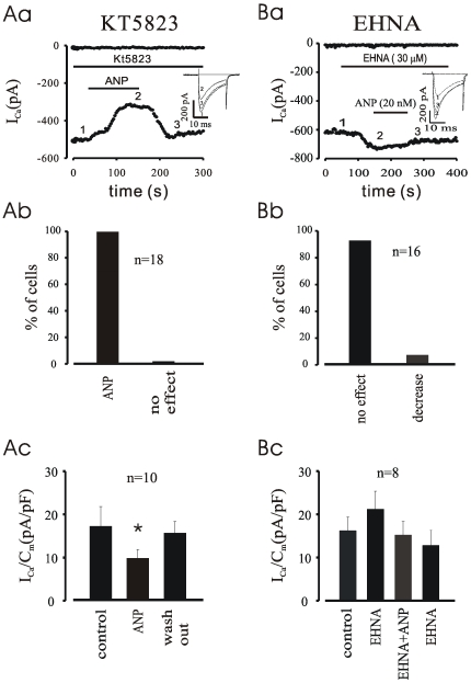
Methodology/principal findings: We investigated the role of ANP in regulating L-type Ca(2+) channel current (I(CaL)) in different developmental stages of cardiomyocytes derived from ES cells. ANP decreased the frequency of action potentials (APs) in early developmental stage (EDS) cardiomyocytes, embryonic bodies (EB) as well as whole embryo hearts. ANP exerted an inhibitory effect on basal I(CaL) in about 70% EDS cardiomyocytes tested but only in about 30% late developmental stage (LDS) cells. However, after stimulation of I(CaL) by isoproterenol (ISO) in LDS cells, ANP inhibited the response in about 70% cells. The depression of I(CaL) induced by ANP was not affected by either Nomega, Nitro-L-Arginine methyl ester (L-NAME), a nitric oxide synthetase (NOS) inhibitor, or KT5823, a cGMP-dependent protein kinase (PKG) selective inhibitor, in either EDS and LDS cells; whereas depression of I(CaL) by ANP was entirely abolished by erythro-9-(2-Hydroxy-3-nonyl) adenine (EHNA), a selective inhibitor of type 2 phosphodiesterase(PDE2) in most cells tested. CONCLUSION/SIGNIFICANCES: Taken together, these results indicate that ANP induced depression of action potentials and I(CaL) is due to activation of particulate guanylyl cyclase (GC), cGMP production and cGMP-activation of PDE2 mediated depression of adenosine 3', 5'-cyclic monophophate (cAMP)-cAMP-dependent protein kinase (PKA) in early cardiomyogenesis.


Conflict of interest statement

Competing Interests: The authors have declared that no competing interests exist.

Figures

Figure 1
Figure 1. Expression of ANP in mouse embryonic cardiomyocytes.
A: double staining of ANP (left, green) and α-MHC (right, red) in E8.5d (a), E12.5d (b), and E15.5d (c) cardiomyocytes. B: PCR product of ANP. Lanes indicate 1 marker, 2 E8.5d, 3 E12.5d, and 4 E15.5d, respectively. The scale bar is 10 micron.
Figure 2
Figure 2. Inhibition of spontaneous action potentials by ANP.
A: Action potentials recorded from a spontaneously contracting EDS cell (upper) and an E8.5d cardiomyocyte (lower) in the current clamp mode. The frequency of action potentials was decreased by application of ANP in a reversible manner. B: action potentials recorded from an early developmental stage EB (upper) and an embryonic 8.5 day heart (lower). As seen in single EDS and E8.5d cells, application of ANP exerted a negative chronotropic effect that was washable.
Figure 3
Figure 3. ANP depresses basal ICaL in EDS cardiomyocytes.
A, Typical recording of ICaL from a single cell. Upon application of 20 nM ANP, rat 3–28, basal ICaL was decreased. This effect could be partially reversed by washout. The inset represents currents recorded as indicated by the mumbers in the time course diagram. B, I/V relationship: ANP decreased peak ICaL without a shift in the I/V relationship. C, percentage of cells responding to ANP. D, density of peak ICaL.
Figure 4
Figure 4. Effect of ANP on basal and ISO stimulated ICaL in LDS cells.
Aa & Ba, time courses of peak ICaL recorded from single LDS cells. Both basal and ISO pre-stimulated ICaL were strongly depressed by application of ANP. Ab & Bb, percentage of cells responding to ANP. Note that the percentage of responding cell in EDS is much lower compared to LDS cells. Ac & Bc, current density of peak ICaL. *P<0.05, **P<0.01 compared with control, respectively.
Figure 5
Figure 5. Effect of ATP-γ-S plus forskolin on ANP induced ICaL inhibition.
Aa, time course shows that ANP depressed forskolin pre-stimulated ICaL, however the depressed effect of ANP on forskolin pre-stimulated ICaL was abolished in the presence of ATP-γ-S (Ba). The insets are traces recorded in the same cells as time courses. Ab & Bb, percentage of cells with or without responding to ANP. Ac & Bc, density of peak ICaL. *P<0.05 compared with control.
Figure 6
Figure 6. PTX did not abolish ANP induced ICaL inhibition.
Aa, time course demonstrates that in PTX pre-treated cardiomyocytes derived from ES cells ANP still inhibited ICaL. Ba, time course of PTX positive control experiment which indicates PTX abolished CCh caused inhibition on ICaL. Ab and Bb,percentage of cells with or without responding to ANP(Ab) or CCh (Bb). Ac and Bc, density of peak ICaL. *P<0.05 compared with control.
Figure 7
Figure 7. PDE2-cAMP-PKA pathway of ANP depression on ICaL.
A, time course of ICaL (a) shows ANP still inhibits the current in KT5823, a PKG selective inhibitor, pre-treated myocytes in about 70% cells tested (b); Ac, density of peak ICaL. B, time course (a) demonstrates that ANP fails to depress ICaL in the presence of EHNA, a PDE2 selective inhibitor, in almost all cells tested (b). Bc, density of peak ICaL. *P<0.05 compared with control.

Similar articles

Cited by

References

    1. Nilius B, Boldt W, Benndorf K. Properties of aconitine-modified sodium channels in single cells of mouse ventricular myocardium. Gen Physiol Biophys. 1986;5:473–484. - PubMed
    1. Kohya T, Tomita F, Itoh K, Suzuki Y, Kawabata N, et al. Silent myocardial ischemia during Holter monitoring in ischemic heart disease. Jpn Circ J. 1989;53:1399–1406. - PubMed
    1. Semmekort B, Guignard JP. Atrial natriuretic peptide during early human development. Biol Neonate. 1991;60:341–349. - PubMed
    1. Brenner BM, Ballermann BJ, Gunning ME, Zeidel ML. Diverse biological actions of atrial natriuretic peptide. Physiol Rev. 1990;70:665–699. - PubMed
    1. Chinkers M, Garbers DL, Chang MS, Lowe DG, Chin HM, et al. A membrane form of guanylate cyclase is an atrial natriuretic peptide receptor. Nature. 1989;338:78–83. - PubMed

MeSH terms