. 2010 Jan 22;5(1):e8854.
doi: 10.1371/journal.pone.0008854.

Pea3 transcription factors and wnt1-induced mouse mammary neoplasia

Affiliations

Pea3 transcription factors and wnt1-induced mouse mammary neoplasia

Rebecca Baker et al. PLoS One. .

Abstract

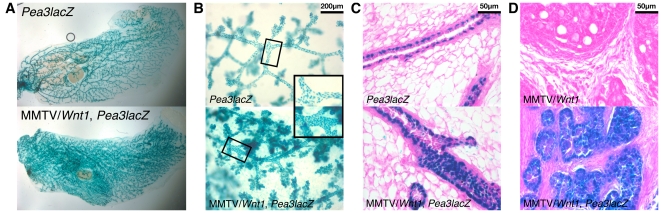
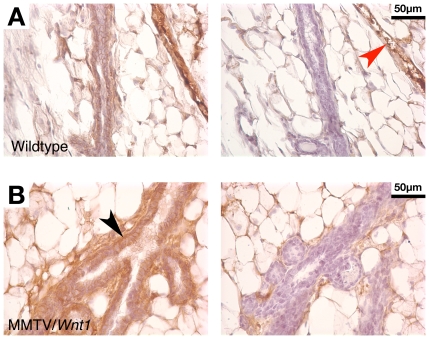
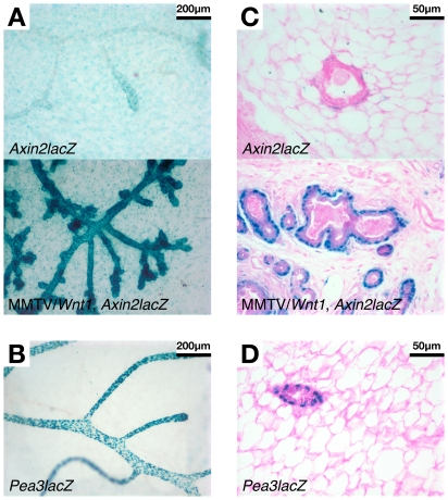
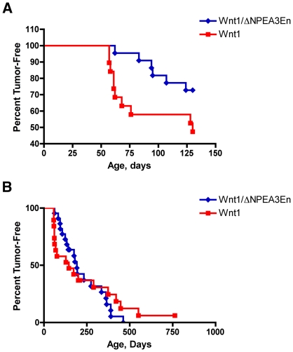
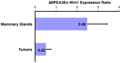
The role of the PEA3 subfamily of Ets transcription factors in breast neoplasia is controversial. Although overexpression of PEA3 (E1AF/ETV4), and of the related factors ERM (ETV5) and ER81 (ETV1), have been observed in human and mouse breast tumors, PEA3 factors have also been ascribed a tumor suppressor function. Here, we utilized the MMTV/Wnt1 mouse strain to further interrogate the role of PEA3 transcription factors in mammary tumorigenesis based on our previous observation that Pea3 is highly expressed in MMTV/Wnt1 mammary tumors. Pea3 expression in mouse mammary tissues was visualized using a Pea3(NLSlacZ) reporter strain. In normal mammary glands, Pea3 expression is predominantly confined to myoepithelial cells. Wnt1 transgene expression induced marked amplification of this cell compartment in nontumorous mammary glands, accompanied by an apparent increase in Pea3 expression. The pattern of Pea3 expression in MMTV/Wnt1 mammary glands recapitulated the cellular profile of activated beta-catenin/TCF signaling, which was visualized using both beta-catenin immunohistochemistry and the beta-catenin/TCF-responsive reporter Axin2(NLSlacZ). To test the requirement for PEA3 factors in Wnt1-induced tumorigenesis, we employed a mammary-targeted dominant negative PEA3 transgene, DeltaNPEA3En. Expression of DeltaNPEA3En delayed early-onset tumor formation in MMTV/Wnt1 virgin females (P = 0.03), suggesting a requirement for PEA3 factor function for Wnt1-driven tumor formation. Consistent with this observation, expression of the DeltaNPEA3En transgene was profoundly reduced in mammary tumors compared to nontumorous mammary glands from bigenic MMTV/Wnt1, MMTV/DeltaNPEA3En mice (P = 0.01). Our data provide the first description of Wnt1-mediated expansion of the Pea3-expressing myoepithelial compartment in nontumorous mammary glands. Consistent with this observation, mammary myoepithelium was selectively responsive to Wnt1. Together these data suggest the MMTV/Wnt1 strain as a potential model of basal breast cancer. Furthermore, this study provides evidence for a protumorigenic role of PEA3 factors in breast neoplasia, and supports targeting the PEA3 transcription factor family in breast cancer.


Conflict of interest statement

Competing Interests: The authors have declared that no competing interests exist.

Figures

Figure 1
Figure 1. The Pea3NLSlacZ strain acts as a reporter of in vivo Pea3 expression in mammary glands.
Abdominal (#4) mammary glands were harvested from female mice that were wildtype (WT), MMTV/Wnt1, Pea3+/NLSlacZ (Pea3lacZ), and bigenic MMTV/Wnt1, Pea3+/NLSlacZ (MMTV/Wnt1, Pea3lacZ). Mammary glands were stained with X-gal as previously described and wholemounted. Staining of epithelial structures was observed in mammary glands from Pea3+/NLSlacZ females and bigenic MMTV/Wnt1, Pea3+/NLSlacZ females, but not in tissues from wildtype or MMTV/Wnt1 animals.
Figure 2
Figure 2. Wnt1 expression in mammary tissues drives expansion of the Pea3-expressing myoepithelial population.
Mammary tissues were harvested from age-matched virgin female littermates and stained with X-gal. (A) Wholemounted glands (10 weeks old)–entire gland. (B) Wholemounted glands (30 weeks old) viewed at 10× magnification (inset, enlargement of boxed area). (C) Mammary gland tissue sections (10 weeks old), counterstained with eosin, viewed at 40× magnification. (D) Tumor tissue sections, counterstained with eosin, viewed at 40× magnification. β-gal activity, indicative of Pea3 expression, was detected primarily in myoepithelial cells in normal mammary glands. The frequency of Pea3-expressing cells was increased in Wnt1-expressing mammary glands, and there was an apparent increase in signal intensity on a per cell basis. Pea3 was widely expressed throughout the tumor epithelium in MMTV/Wnt1 mammary tumors.
Figure 3
Figure 3. Wnt1 expression in mouse mammary glands induces profound β-catenin accumulation in the myoepithelial compartment.
Mammary gland tissue sections from virgin female mice (10 weeks old) were subjected to β-catenin IHC as previously described and counterstained with hematoxylin. As a control, serial sections were stained in parallel omitting primary antibody (right-hand panels). (A) Wildtype. (B) MMTV/Wnt1. Weak staining of membrane-localized β-catenin was observed in wildtype epithelium. Vascular structures were clearly evident in control slides stained with secondary antibody alone (red arrowhead). Profound increases in β-catenin protein levels were observed in MMTV/Wnt1 epithelium, including both increased membrane signal as well as β-catenin accumulation in the cytoplasm and nucleus. Cells in the myoepithelial compartment exhibited the strongest β-catenin signal (black arrowhead).
Figure 4
Figure 4. β-catenin/TCF signaling activity revealed using Axin2NLSlacZ mice mirrors nucleocytoplasmic β-catenin accumulation.
Abdominal (#4) mammary glands were harvested from female mice that were Axin2+/NLSlacZ (Axin2lacZ) or bigenic MMTV/Wnt1, Axin2+/NLSlacZ (MMTV/Wnt1, Axin2lacZ), and were stained with X-gal as previously described . Pea3+/NLSlacZ (Pea3lacZ) samples were also included for comparison. (A) Wholemounted glands (11 weeks old), viewed at 10× magnification. (B) Wholemounted gland from Pea3lacZ virgin female stained in parallel with the specimens in Panel A, viewed at 10× magnification. (C) Mammary gland tissue sections (11 weeks old), counterstained with eosin, viewed at 40× magnification. (D) Mammary gland tissue section from Pea3lacZ virgin female (10 weeks old), counterstained with eosin, viewed at 40× magnification. β-gal activity was detected in adipocytes throughout the mammary fat pad from both Axin2+/NLSlacZ and bigenic MMTV/Wnt1, Axin2+/NLSlacZ mice. Additionally, in Axin2+/NLSlacZ mammary gland, cells in the connective tissue peripheral to the mammary epithelium stained positive, but no staining was detected in the myoepithelial or luminal cells. Wnt1 expression induced robust β-gal activity, indicative of β-catenin/TCF signaling, in the myoepithelial compartment, with weaker but detectable signal in the luminal epithelium.
Figure 5
Figure 5. The dominant-interfering ΔNPEA3En transgene suppresses early onset tumor formation in MMTV/Wnt1 mice.
Tumor latency was compared in two cohorts of virgin female mice, MMTV/Wnt1 (red squares; n = 19) and bigenic MMTV/Wnt1, MMTV/ΔNPEA3En (blue diamonds; n = 22), generated by interbreeding MMTV/Wnt1 males with MMTV/ΔNPEA3En females. (A) Formation of early onset tumors was substantially retarded by expression of the dominant-interfering ΔNPEA3En transgene (P = 0.03, log-rank test). Survival data were censored at 130 days to compare early onset tumor formation (B) The overall rate of tumor formation was similar in both cohorts, suggesting that tumors were ultimately able to escape from ΔNPEA3En-mediated tumor suppression (P = 0.39, log-rank test).
Figure 6
Figure 6. ΔNPEA3En expression is markedly decreased in tumors relative to mammary glands from MMTV/Wnt1, MMTV/ΔNPEA3En mice.
Expression levels of the Wnt1 and ΔNPEA3En transgenes in mammary glands and tumors from bigenic MMTV/Wnt1, MMTV/ΔNPEA3En mice were assayed by RT-PCR. The expression level of ΔNPEA3En in each sample was normalized to that of Wnt1 in the same sample to control for potential variations in epithelial content between tumors and nontumorous mammary glands. The ratio of ΔNPEA3En expression to Wnt1 expression was strikingly reduced in mammary tumors relative to non-tumorous mammary glands (P = 0.01, t-Test).

Similar articles

Cited by

References

    1. Graves BJ, Petersen JM. Specificity within the ets family of transcription factors. Adv Cancer Res. 1998;75:1–55. - PubMed
    1. Sharrocks AD. The ETS-domain transcription factor family. Nat Rev Mol Cell Biol. 2001;2:827–837. - PubMed
    1. Shepherd T, Hassell JA. Role of Ets transcription factors in mammary gland development and oncogenesis. J Mammary Gland Biol Neoplasia. 2001;6:129–140. - PubMed
    1. Martin ME, Piette J, Yaniv M, Tang WJ, Folk WR. Activation of the polyomavirus enhancer by a murine activator protein 1 (AP1) homolog and two contiguous proteins. Proc Natl Acad Sci U S A. 1988;85:5839–5843. - PMC - PubMed
    1. Wasylyk C, Flores P, Gutman A, Wasylyk B. PEA3 is a nuclear target for transcription activation by non-nuclear oncogenes. Embo J. 1989;8:3371–3378. - PMC - PubMed

Publication types

LinkOut - more resources