. 2010 Jan 22;5(1):e8843.
doi: 10.1371/journal.pone.0008843.

Temporal and spatial requirements of unplugged/MuSK function during zebrafish neuromuscular development

Affiliations

Temporal and spatial requirements of unplugged/MuSK function during zebrafish neuromuscular development

Lili Jing et al. PLoS One. .

Abstract

One of the earliest events in neuromuscular junction (NMJ) development is the accumulation of acetylcholine receptor (AChR) at the center of muscle cells. The unplugged/MuSK (muscle specific tyrosine kinase) gene is essential to initiate AChR clustering but also to restrict approaching growth cones to the muscle center, thereby coordinating pre- and postsynaptic development. To determine how unplugged/MuSK signaling coordinates these two processes, we examined the temporal and spatial requirements of unplugged/MuSK in zebrafish embryos using heat-shock inducible transgenes. Here, we show that despite its expression in muscle cells from the time they differentiate, unplugged/MuSK activity is first required just prior to the appearance of AChR clusters to simultaneously induce AChR accumulation and to guide motor axons. Furthermore, we demonstrate that ectopic expression of unplugged/MuSK throughout the muscle membrane results in wildtype-like AChR prepattern and neuromuscular synapses in the central region of muscle cells. We propose that AChR prepatterning and axonal guidance are spatio-temporally coordinated through common unplugged/MuSK signals, and that additional factor(s) restrict unplugged/MuSK signaling to a central muscle zone critical for establishing mid-muscle synaptogenesis.


Conflict of interest statement

Competing Interests: The authors have declared that no competing interests exist.

Figures

Figure 1
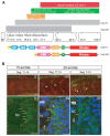
Figure 1. Neuromuscular synaptogenesis in zebrafish.
(A) Schematic representation of the different steps during neuromuscular synapse development. unplugged SV1 mRNA is expressed transiently from the tailbud stage to 48 hpf, while unplugged FL mRNA is expressed from the 10-somite stage and continues to be expressed in adult muscle. Bottom: Domain structure of the unplugged FL and SV1 isoforms. (B) Synaptogenesis at the 15- or 20-somite stage. 15-somite (B1) or 20-somite stage (B3 and B4) embryos stained for motor axons (znp-1, green) and AChR clusters (α-BTX, red). In segments 11–14, AChR prepatterned clusters appear at the 15-somite stage before motor neurons have exited the spinal cord (B1); prepatterned clusters coalesce and motor axons (arrows) begin to approach the myotome at the 20-somite stage (B3). In anterior older segments 7–10, motor axons have contacted non-migratory adaxial cells (arrowheads) at the 20-somite stage (B5). (B2, B4, B6) Cross-sectional views of Tg(smyhc1:mcherry-CAAX) embryos, which express mCherry specifically on the membrane of adaxial cells, stained for adaxial muscles (anti-DsRed, green), prepatterned clusters (α-BTX, red) and nuclei (DAPI, blue). The same segments as in (B1, B3, B5) were analyzed. (B2) At the 15-somite stage, adaxial muscles remain adjacent to notochord, and prepatterned clusters are localized at the medial side of the cells (small arrows). (B4) At the 20-somite stage, adaxial muscles have migrated laterally. Some prepatterned clusters co-migrate with the adaxial cells (big arrow). Prepatterned clusters appear on the medial side of fast muscle fibers (arrow heads). Prepatterned clusters remain on the medial side of the non-migratory adaxial cells (small arrows). (B6) In older segments 7–10, adaxial muscles have migrated further away; AChR clusters remain on the medial side of non-migratory adaxial cells (small arrows). AChR clusters appear on the medial side of fast muscle fibers (arrow heads). NC: notochord. Scale bars: 20 µM in B; 10 µM in B2 and B4.
Figure 2
Figure 2. Transgene expression after heat-shock (HS) induction.
(A) Lateral views of 26 hpf whole-mount embryos stained with anti znp1 (notor axons, green) and anti myc (red) antibodies. No Myc protein expression is detectable in non-transgenic wildtype and transgenic embryos, but is ubiquitously detectable 10 minutes after a 40-min HS treatment in transgenic embryos. HS treatment was performed at 24hpf for 40 minutes, embryos were fixed and processed at 26 hpf. (B) unplugged/MuSK-myc fusion protein was visualized by western blot with an anti-myc antibody (1:500) in non-transgenic wildtype, transgenic non-heat shocked controls, and 30 minutes, 3 hours, 6 hours and 9 hours after a 40-min HS induction of Tg(hsp70l:unplugged SV1-myc). An anti-actin antibody (1∶500) is used as a loading control. Tg(hsp70l:unplugged FL-myc) shows similar results (data not shown). HS treatment was performed at 24hpf for 40 minutes, embryos were fixed and processed at indicated times post heat shock.
Figure 3
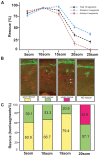
Figure 3. Rescue of motor axon pathfinding and AChR prepatterning by unplugged/MuSK at different time points.
(A) Temporal series of Tg(hsp70l:unpFLmyc) induction to rescue motor axon guidance. Embryos were heat-shocked starting at the indicated time points and analyzed at 27 hpf. The induction of Tg(hsp70l:unpFLmyc) expression after the 15-somite stage significantly reduced rescue efficacy (segments 6–15, black; segments 6–10, red; segments 11–15, blue). See Materials and Methods for how rescue efficiency was calculated. 20 hemisegments were analyzed in each embryo, and results from multiple experiments are represented as mean±SEM. (n = 644−1028, average = 744, hemisegments per time point). (B and C) Rescue of AChR prepatterning by Tg(hsp70l:unplugged SV1-myc). Embryos were heat-shocked starting at different time points and examined at the 21-somite stage for prepatterned AChR clusters. Only posterior segments (11–15) were analyzed in each embryo (see also Figure S3). (B) Individual hemisegments were scored as ‘good rescue’ (when AChR clusters were present on most muscles), ‘mild rescue’ (when AChR clusters formed on <50% of all muscles, mostly around non-migratory adaxial cells), or ‘weak rescue’ (with smaller prepatterned clusters formed on a few muscles, see arrows). (C) HS treatment of Tg(hsp70l:unplugged SV1-myc) embryos at the 20-somite stage significantly reduced AChR rescue efficacy. Results were summarized from multiple experiments (n = 7−20, average = 13, hemisegments per time point). Scale bar: 20 µM.
Figure 4
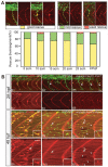
Figure 4. Rescue of neuromuscular synapses by unplugged/MuSK.
(A) Rescue of neural synapses at different induction times by Tg(hsp70l:unplugged FL-myc). Embryos were heat-shocked starting at the indicated time points, fixed at 27hpf, and stained for motor axons (znp-1, green) and AChR clusters (α-BTX, red). Individual hemisegments were scored as ‘good rescue’ (when AChR clusters colocalized with presynaptic nerve ending on most muscle fibers, including along ectopic nerve ending), ‘mild rescue’ (when synapses formed on <50% of all muscles), or ‘weak rescue’ (when synapses formed only on non-migratory adaxial cells). 20 hemisegments were scored in each embryo. Results were summarized from two independent experiments (n = 160−380, average = 280, hemisegments per time point). (B) Tg(hsp70l:unplugged FL-myc) rescues neuromuscular synapses at 28 hpf and 48 hpf. Embryos were given a 40-min HS at 28hpf or 48hpf, and examined 30 minutes after the HS treatment. (B1–B6) In transgenic embryos, neuromuscular synapses formed on most muscle fibers and aligned with nerve ending. Arrows point to unplugged-characteristic pathfinding errors, indicating that the HS was too late to rescue axon pathfinding. (B7–B12) The single HS treatment at 48 hpf induced synapses in transgenic embryos (arrowheads in B11 and B12), albeit synapses appear smaller than those in wildtype. Inset in each panel is the enlarged image of the neuromuscular synapse pointed by the arrow. Scale bars: 20 µM.
Figure 5
Figure 5. Overexpression of unplugged/MuSK in WT embryos does not perturb synaptogenesis.
21-somite stage (A–F) or 27 hpf (G–L) embryos after HS treatment. (A–F) Wildtype (WT) embryos, Tg(hsp70l:unplugged SV-1myc) embryos, and Tg(hsp70:unplugged SV1-myc); unpluggedbr307/br307 embryos received HS from the 10- to 21-somite stage The AChR prepatterning zone appears slightly expanded in (D) compared to (B and F). White dashed lines mark the boundaries of each segment. (G–L) WT embryos, hsp70l:unplugged SV-myc embryos, (I and J) or hsp70l:unplugged FL-myc embryos (K and L) were heat shocked from the 10-somite stage until 27 hpf. Motor axons and neuromuscular synapses appear normal in the embryos expressing the transgene (I–L). Scale bars: 20 µM.
Figure 6
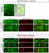
Figure 6. Cellular localization of exogenous unplugged/MuSK protein and AChR clusters.
(A) Tg(hsp70l:unplugged SV1-myc); unplugged br307/br307 embryos at the 10-somite stage after the HS treatment, stained for anti-Myc Ab (green). Prior to the appearance of AChR clusters Unplugged SV1 protein is distributed evenly on the surface of adaxial muscle cells. (B) Diagram of A. (C–H) Tg(hsp70l:unplugged SV1-myc); unplugged br307/br307 embryos at the 21-somite stage after the HS treatment, stained for anti-Myc Ab (green) and AChR clusters (α-BTX, red). Lateral views (C–E) and cross-sectional views (F–H) show that Unplugged SV1 protein is distributed throughout the surface of muscle cells, but prepatterned clusters are localized at the center and the medial side of the cell surface. (I–Q) Tg(hsp70l:unplugged FL-myc); unplugged br307/br307 embryos at 27 hpf after HS treatment, stained for anti-Myc Ab (green, I–Q) and AChR clusters (α-BTX, red, J,K,M,N) or znp1(red, P–Q). Unplugged FL protein is expressed throughout the cell membrane as shown in lateral views (I–K), or cross-sectional views, (L–Q). Neural AChR clusters and motor axons are located at the center and the medial side of the cell. Non-migratory adaxial cells at the horizontal myoseptum were imaged. White dashed lines mark the boundaries of the somite in (C–E, I–K) or indicate the position of notochord in (A, B, F–H, L–Q). NC, notochord. Scale bars: 10 µM.

References

    1. Sanes JR, Lichtman JW. Induction, assembly, maturation and maintenance of a postsynaptic apparatus. Nat Rev Neurosci. 2001;2:791–805. - PubMed
    1. Kummer TT, Misgeld T, Sanes JR. Assembly of the postsynaptic membrane at the neuromuscular junction: paradigm lost. Curr Opin Neurobiol. 2006;16:74–82. - PubMed
    1. Witzemann V. Development of the neuromuscular junction. Cell Tissue Res. 2006;326:263–271. - PubMed
    1. Flanagan-Steet H, Fox MA, Meyer D, Sanes JR. Neuromuscular synapses can form in vivo by incorporation of initially aneural postsynaptic specializations. Development 2005 - PubMed
    1. Panzer JA, Song Y, Balice-Gordon RJ. In vivo imaging of preferential motor axon outgrowth to and synaptogenesis at prepatterned acetylcholine receptor clusters in embryonic zebrafish skeletal muscle. J Neurosci. 2006;26:934–947. - PMC - PubMed

Publication types

MeSH terms

LinkOut - more resources