. 2010 Jan 22;5(1):e8846.
doi: 10.1371/journal.pone.0008846.

TBCD links centriologenesis, spindle microtubule dynamics, and midbody abscission in human cells

Affiliations

TBCD links centriologenesis, spindle microtubule dynamics, and midbody abscission in human cells

Mónica López Fanarraga et al. PLoS One. .

Abstract

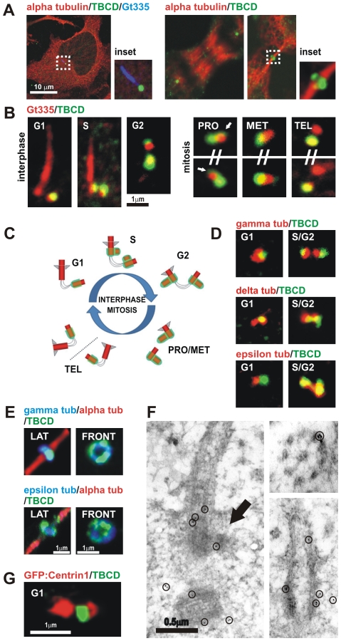
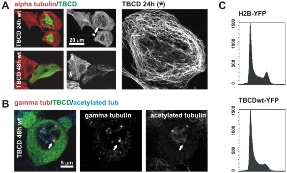
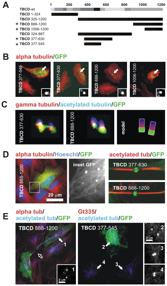
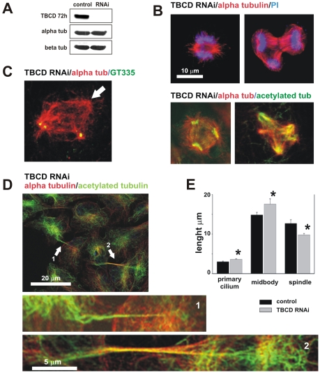
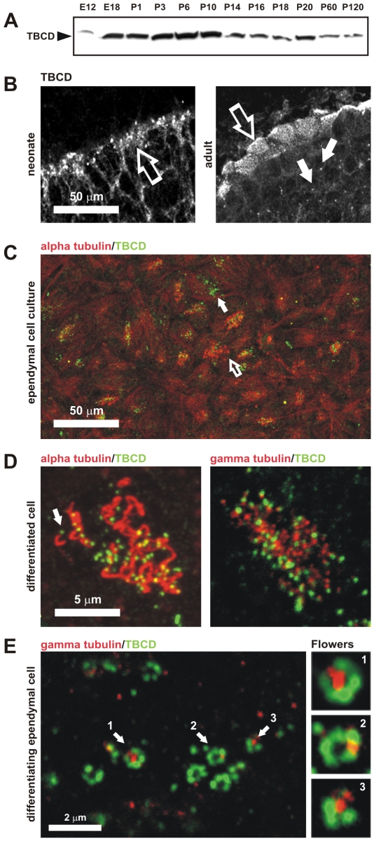
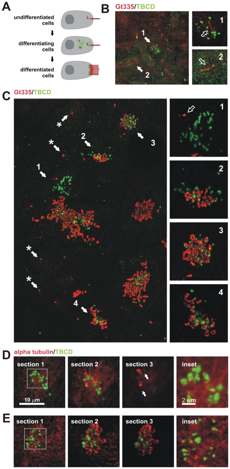
Microtubule-organizing centers recruit alpha- and beta-tubulin polypeptides for microtubule nucleation. Tubulin synthesis is complex, requiring five specific cofactors, designated tubulin cofactors (TBCs) A-E, which contribute to various aspects of microtubule dynamics in vivo. Here, we show that tubulin cofactor D (TBCD) is concentrated at the centrosome and midbody, where it participates in centriologenesis, spindle organization, and cell abscission. TBCD exhibits a cell-cycle-specific pattern, localizing on the daughter centriole at G1 and on procentrioles by S, and disappearing from older centrioles at telophase as the protein is recruited to the midbody. Our data show that TBCD overexpression results in microtubule release from the centrosome and G1 arrest, whereas its depletion produces mitotic aberrations and incomplete microtubule retraction at the midbody during cytokinesis. TBCD is recruited to the centriole replication site at the onset of the centrosome duplication cycle. A role in centriologenesis is further supported in differentiating ciliated cells, where TBCD is organized into "centriolar rosettes". These data suggest that TBCD participates in both canonical and de novo centriolar assembly pathways.


Conflict of interest statement

Competing Interests: The authors have declared that no competing interests exist.

Figures

Figure 1
Figure 1. TBCD is concentrated in centrioles and midbodies.
(A) Confocal-microscopic images of a HeLa cell (ATCCR number: CCL-2™) immunostained for tubulin, TBCD, and GT335 (which labels glutamylated tubulin at the centrioles and primary cilium), where a single spot of TBCD is observed close to the basal body of the cilium. HeLa cells in anaphase and undergoing cytokinesis display clear centrosomal labelling and recruitment of TBCD to the midbody ring, respectively. (B) High-resolution confocal-microscopic section images of doubly immunostained centrosomes of HeLa cells. The cells were photographed at different stages of the cell cycle, labelled with GT335 and anti-TBCD antibody. This tubulin cofactor is observed at one of the centrioles during G1. Two stronger additional TBCD signals immediately adjacent to the existing centrioles, devoid of GT335 staining, were stained at G1. By late S three TBCD spots were detected. By prophase, most dividing cells contained two strong TBCD spots, one in each centrosome. Partial TBCD halos surrounding older centrioles were also observed (arrows). At the end of telophase, only one of the centrioles in each daughter cell was labelled for TBCD. (C) Diagram of the distribution of TBCD during the centriolar cycle; centrioles, procentrioles, and primary cilium are shown in red and TBCD is shown in green. (D) Partial co-localization of the TBCD signal with γ- and δ-tubulins was observed throughout the cell cycle. The ε-tubulin signal concentrated at the mother centriole during G1 did not co-localize with TBCD. Later in the cell cycle, both proteins appeared to partially co-localize. (E) TBCD accumulated at the midbody, where γ- and ε-tubulins were also detected. Lateral and frontal views of the structure correspond to different cells. (F) Immuno-electron-microscopic analysis of TBCD on murine epithelial cells. (Left) TBCD localises on the proximal region of the basal body (the original mother centriole) during procentriole assembly (arrow) S stage. The former daughter centriole is also observed in the section (gold particles are outlined with circles). (Right top) TBCD labelling was also detected at the proximal ends of basal bodies and (Right bottom) at the outer microtubule doublets of motile tracheal cilia. (G) Immunostaining of HeLa cells transfected with GFP-centrin1 (red, pseudocolor) confirm TBCD localization at the proximal end of the daughter centriole.
Figure 2
Figure 2. TBCD overexpression resulted in microtubule detachment from the centrosome and spindle abnormalities.
(A) Confocal-microscopic projected images of a HeLa cell overexpressing HsTBCD doubly immunostained for tubulin and TBCD. (Top) Microtubule release from the centrosome was clear in most cells 15 h after transfection (arrow). (Bottom) Microtubule network destruction occurred approximately 30 h after transfection. Right panel shows a closer view of the microtubule pattern of the cell labelled with an arrow.(B) TBCD overexpression also produced aberrant mitotic figures, where acentriolar supernumerary MTOCs containing γ-tubulin were observed (arrow). (C) Flow-cytometric profiles of cells (>10,000 cells per profile) labelled with Hoechst and transfected with HsTBCD–YFP or a control–YFP construct. Clear G1 arrest, typical of the loss of centrosomal integrity, was observed in the cells analysed at both 15 and 30 h after transfection.
Figure 3
Figure 3. TBCD contains a microtubule-binding region and two centriolar-targeting regions.
(A) Diagram of the full-length TBCD polypeptide and the truncation mutants produced for this study. The degree of evolutionary conservation of the polypeptide is shown in relative shades of grey. (B) Confocal images of HeLa cells transfected with constructs encoding GFP-fusion TBCD truncation mutants and immunostained for tubulin. The constructs shown correspond to those labelled with an asterisk in A. The GFP labelling at the centrosomal region is shown in the inset. (C) High-resolution confocal images of triply labelled centrioles in weakly expressing cells show the co-localization of these two TBCD truncation mutants with γ-tubulin in the proximal region of both centrioles. (D) TBCD contains a microtubule-binding region. GFP–TBCD888–1200-decorated microtubules 48 h after transfection. (Left) TBCD centrosomal-binding truncation mutants were also recruited to the Fleming bodies of cells at the end of mitosis. (E) Phenotypes observed for TBCD centrosomal-binding truncation mutants. Aberrant midbodies with increased lengths and reduced thicknesses were common (right, open arrow). Centriolar separation was increased (>2 µm) in many of the transfected cells (centrosomes 1, 2 versus 3, 4).
Figure 4
Figure 4. TBCD depletion resulted in microtubule spindle abnormalities and failure of cell abscission.
(A) Western blot confirmation of TBCD silencing 72 h after siRNA treatment. Whole cell lysates (50 µg/lane) were loaded and analysed by immunoblotting with antibodies directed against CoD and the α- and β-tubulins. TBCD depletion did not affect α- or β-tubulin levels. (B) Confocal-microscopic projected images of different mitotic spindle defects observed after TBCD interference in HeLa cell cultures. Mitotic aberrations included abnormally short spindles, abnormal anaphase figures, and multipolar spindle defects, among others. (C) Acentriolar spindle poles were also observed (arrow). (D) TBCD silencing resulted in cell abscission failure. The maintenance of cytoplasmic bridges containing microtubules is shown (1, 2). These abnormally long midbodies are characterized by their content of acetylated microtubules (2). (E) Statistical analysis of the length of the primary cilia (DF = 147; P = 3×10⁻³) and midbodies (DF = 174; P = 0.1) confirmed significant increases in the lengths of these two structures after TBCD depletion. A highly significant reduction in the spindle pole-to-pole distance (DF = 63; P = 5×10⁻⁴) was also detected (*).
Figure 5
Figure 5. TBCD is required in ciliogenesis.
(A) TBCD is abundantly expressed during neurogenesis. Western blot of total protein extracts (50 µg/lane) from developing and adult mouse brains obtained at the indicated embryonic and postnatal ages. (B) Confocal-microscopic images of cryo-sections of neonatal and adult murine cerebral ependymal epithelium immunostained for TBCD. This cofactor accumulated in clusters localized in the ependymal cell cytoplasm (left, open arrow). In the adult, TBCD localized at the apical borders of the ependymal cells, where the basal bodies are located (right, open arrow). TBCD was also detectable on parenchymal cell centrosomes (solid arrow). (C) A general view of an ependymal cell primary culture in which cilia, immunostained with anti-tubulin antibody, and TBCD are labelled. These culture conditions allow the observation of epithelial cells at various differentiation stages (I–IV, see the text). (Solid arrow) TBCD accumulated in differentiating cells with no cilia, whereas ciliated cells (open arrow) contained fewer TBCD spots. (D) Detail of the apical borders of differentiated ependymal cells where a single TBCD spot was observed at the base of each cilium (arrow). These TBCD spots partially co-localized with the γ-tubulin-labelled basal body. (E) Detail of the cytoplasm of a differentiating ependymal cell (stages I–II), where the assembly of several centriolar rosettes, labelled for TBCD and γ-tubulin, are shown. TBCD accumulated into structures of approximately 0.3 µm in diameter, which created ring-like structures that were often associated with a single γ-tubulin spot of a similar diameter (1, 2, and 3).
Figure 6
Figure 6. TBCD changes during ciliogenesis in ependymal cells.
(A) Diagram of the location of TBCD accumulation (green) with respect to the centrioles and cilia (red) during ependymal cell differentiation. (B) Short-term cultures display cells at stages I and II, where only a single cilium per cell is observed, presumably the primary cilium. (1) The presence of the primary cilium is often accompanied by the development of small cytoplasmic TBCD clusters containing 1–3 TBCD spots of approximately 0.3 µm. (2) Uncommitted epithelial cells in the culture displayed diffuse TBCD immunostaining. This confocal plane did not coincide with the daughter centriole. (C) General view of a culture immunostained with GT335 and for TBCD. Here, the relationship between TBCD and the assembly of ependymal cilia can be seen. (*) Primary cilia with their corresponding TBCD-labelled centrioles are observed. (1) Differentiating cells (stages I–II) contain TBCD aggregates in their cytoplasm (Figure 5E). (2–4) Cilia assembly occurs after TBCD spots migrate to the apical border of the cells as single spots, presumably accompanying newly developed centrioles. (D-E) A series of confocal sections obtained from the apical borders of the epithelial cells towards the cilia. Cells were immunostained with anti-α-tubulin and anti-TBCD antibodies to show the relationships between this cofactor, the microtubule cytoskeleton, and the cilia. (D) Differentiating cells displaying a few cilia (arrows) show TBCD clusters visible in the cytoplasmic confocal planes (inset). (E) Differentiated cells containing several cilia show smaller TBCD aggregates in the confocal planes, which coincide with the basal bodies of existing cilia (inset).

Similar articles

Cited by

References

    1. Andersen JS, Wilkinson CJ, Mayor T, Mortensen P, Nigg EA, et al. Proteomic characterization of the human centrosome by protein correlation profiling. Nature. 2003;426:570–574. - PubMed
    1. Lewis SA, Tian G, Cowan NJ. The alpha- and beta-tubulin folding pathways. Trends Cell Biol. 1997;12:479–484. - PubMed
    1. López-Fanarraga M, Avila J, Guasch A, Coll M, Zabala JC. Review: postchaperonin tubulin folding cofactors and their role in microtubule dynamics. J Struct Biol. 2001;135:219–229. - PubMed
    1. Szymanski D. Tubulin folding cofactors: half a dozen for a dimer. Curr Biol. 2002;12:767–769. - PubMed
    1. Tian G, Lewis SA, Feierbach B, Stearns T, Rommelaere H, et al. Tubulin subunits exist in an activated conformational state generated and maintained by protein cofactors. J Cell Biol. 1997;138:821–832. - PMC - PubMed

Publication types

Substances