. 2010 Jan 22;6(1):e1000736.
doi: 10.1371/journal.ppat.1000736.

Protease-sensitive synthetic prions

Affiliations

Protease-sensitive synthetic prions

David W Colby et al. PLoS Pathog. .

Abstract

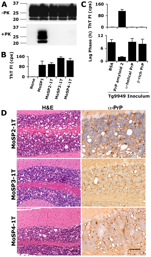
Prions arise when the cellular prion protein (PrP(C)) undergoes a self-propagating conformational change; the resulting infectious conformer is designated PrP(Sc). Frequently, PrP(Sc) is protease-resistant but protease-sensitive (s) prions have been isolated in humans and other animals. We report here that protease-sensitive, synthetic prions were generated in vitro during polymerization of recombinant (rec) PrP into amyloid fibers. In 22 independent experiments, recPrP amyloid preparations, but not recPrP monomers or oligomers, transmitted disease to transgenic mice (n = 164), denoted Tg9949 mice, that overexpress N-terminally truncated PrP. Tg9949 control mice (n = 174) did not spontaneously generate prions although they were prone to late-onset spontaneous neurological dysfunction. When synthetic prion isolates from infected Tg9949 mice were serially transmitted in the same line of mice, they exhibited sPrP(Sc) and caused neurodegeneration. Interestingly, these protease-sensitive prions did not shorten the life span of Tg9949 mice despite causing extensive neurodegeneration. We inoculated three synthetic prion isolates into Tg4053 mice that overexpress full-length PrP; Tg4053 mice are not prone to developing spontaneous neurological dysfunction. The synthetic prion isolates caused disease in 600-750 days in Tg4053 mice, which exhibited sPrP(Sc). These novel synthetic prions demonstrate that conformational changes in wild-type PrP can produce mouse prions composed exclusively of sPrP(Sc).


Conflict of interest statement

The authors have declared that no competing interests exist.

Figures

Figure 1
Figure 1. Tg9949 mice are prone to neurological dysfunction but do not spontaneously generate prions.
(A) Wild-type (FVB), Tg4053, and Tg9949 mice were monitored for signs of neurological dysfunction for >900 days. Both uninoculated and BSA-inoculated Tg9949 mice were more likely than wt mice (p = 0.03) and Tg4053 mice (p<0.001) to develop neurological dysfunction. However, prions could not be detected in the brains of neurologically impaired Tg9949 mice, as determined by bioassay in Tg9949 and Tg4053 mice (B), by histopathological staining (C), by Western immunoblotting (D), and by the amyloid seeding assay (E). (C) Brain sections of neurologically impaired Tg9949 mice were stained with H&E to visualize vacuoles (left), α-GFAP to visualized astrocytic gliosis (middle), and α-PrP to visualize PrPSc deposits (right). Scale bars represent 100 µm. Mol, molecular cell layer; GC, granular cell layer; WM, white matter. Additional neuropathological analyses of control brains are shown in Fig. S1. (D) Western immunoblots of undigested and PK-digested brain samples from six aged Tg9949 mice show no rPrPSc. Homogenate from a Tg9949 mouse inoculated with RML prions is shown as a positive control. Lane assignments are as indicated in panel E. (E) Brain samples from six aged Tg9949 mice do not seed the formation of recPrP amyloid, as judged by an increase in Thioflavin T fluorescence (top) and by a decrease in the lag phase for amyloid formation (bottom). An uninoculated FVB mouse brain is included as a negative control. Negative controls in (A) are pooled results, including some previously published data .
Figure 2
Figure 2. Inoculation of Tg9949 mice with PrP amyloid, but not other PrP conformations, results in the generation of prions.
Tg9949 mice were inoculated with recPrP folded in various conformations (α-helical, β-rich, and amyloid) and allowed to live out their normal life span (Table S2 and S3). Tg9949 mice inoculated with BSA or MoSP1 are shown as controls. The first transmission (1T) of MoSP2, MoSP3, and MoSP4 refers to inoculation of Tg9949 mice with amyloid 2, amyloid 3, and amyloid 4 preparations, respectively. Brain samples from Tg9949 mice containing MoSP2, MoSP3, or MoSP4 did not harbor protease-resistant PrP (A) but showed activity in the ASA (B; kinetic data shown in Fig. S2; note that the sample amount for ASA is 1/1000 of the amount used in Western blots). Brain samples from mice inoculated with non-amyloid PrP conformations did not show activity in the ASA (C). Activity in the ASA was detected by an increase in ThT fluorescence (B and top of panel C) and by a decrease in the mean lag phase for PrP amyloid formation (bottom of panel C). (D) Brain sections of Tg9949 mice containing MoSP2, MoSP3, or MoSP4 showed neuropathology consistent with prion disease, including extensive vacuolation (H&E stain, left column) and PrP deposition (anti-PrP stain, right column). Each scale bar represents 100 µm and applies to the micrographs in the same column.
Figure 3
Figure 3. Protease-sensitive synthetic prions are serially transmissible in Tg9949 mice.
MoSP2-1T, MoSP3-1T and MoSP4-1T were serially passaged to new groups of Tg9949 mice for a second transmission (2T) by intracerebral inoculation of brain homogenate containing each isolate. MoSP2-2T was serially passaged an additional time for a third transmission (3T). In each case, the animals lived a normal life span (Table S4). The brains of Tg9949 mice containing MoSP2-2T, MoSP3-2T, or MoSP4-2T showed no protease-resistant PrP by Western blotting (A), but activity in the ASA (B) and neuropathology consistent with prion disease (C). Brain samples from mice inoculated either with MoSP1 or with homogenates of uninfected Tg9949 mice are shown as controls. (C) Cerebellar sections were stained with H&E (top row) and α-PrP (bottom row). m, molecular layer; gc, granule cell layer. Each scale bar represents 100 µm and applies to the panels in the same row.
Figure 4
Figure 4. Protease-sensitive synthetic prions are serially transmissible to Tg4053 mice.
Tg4053 mice intracerebrally inoculated with MoSP2-1T (red), MoSP19A (blue) or MoSP19B (purple) developed signs of prion disease between 600–750 d (A). MoSP19A and MoSP19B are two Tg9949 brain isolates inoculated with Amyloid Prep 19 (origin in Table S3). Tg4053 mice inoculated with protease-sensitive synthetic prions were significantly more likely to develop neurological dysfunction than mice inoculated with uninfected Tg9949 brain homogenate (black, p<0.001). PrP in the brains of these mice was sensitive to PK digestion (B) and active in the ASA (C). MoSP1 was used as a control. The brains of ill, MoSP2-inoculated Tg4053 mice showed neuropathology consistent with prion disease (D), including vacuolation (top panel), astrocytic gliosis (middle panel), and punctate PrP deposits (bottom panel). Scale bars represent 100 µm.

Similar articles

Cited by

References

    1. Prusiner SB. Prions. Proc Natl Acad Sci USA. 1998;95:13363–13383. - PMC - PubMed
    1. Oesch B, Westaway D, Wälchli M, McKinley MP, Kent SBH, et al. A cellular gene encodes scrapie PrP 27–30 protein. Cell. 1985;40:735–746. - PubMed
    1. Prusiner SB. Novel proteinaceous infectious particles cause scrapie. Science. 1982;216:136–144. - PubMed
    1. McKinley MP, Bolton DC, Prusiner SB. A protease-resistant protein is a structural component of the scrapie prion. Cell. 1983;35:57–62. - PubMed
    1. Meyer RK, McKinley MP, Bowman KA, Braunfeld MB, Barry RA, et al. Separation and properties of cellular and scrapie prion proteins. Proc Natl Acad Sci USA. 1986;83:2310–2314. - PMC - PubMed

Publication types